. 2011 Mar 24;6(3):e18002.
doi: 10.1371/journal.pone.0018002.

Distinct roles of Cdc42 in thymopoiesis and effector and memory T cell differentiation

Affiliations

Distinct roles of Cdc42 in thymopoiesis and effector and memory T cell differentiation

Fukun Guo et al. PLoS One. .

Abstract

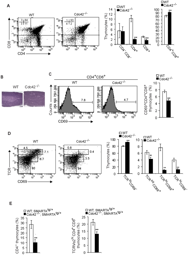
Cdc42 of the Rho GTPase family has been implicated in cell actin organization, proliferation, survival, and migration but its physiological role is likely cell-type specific. By a T cell-specific deletion of Cdc42 in mouse, we have recently shown that Cdc42 maintains naïve T cell homeostasis through promoting cell survival and suppressing T cell activation. Here we have further investigated the involvement of Cdc42 in multiple stages of T cell differentiation. We found that in Cdc42(-/-) thymus, positive selection of CD4(+)CD8(+) double-positive thymocytes was defective, CD4(+) and CD8(+) single-positive thymocytes were impaired in migration and showed an increase in cell apoptosis triggered by anti-CD3/-CD28 antibodies, and thymocytes were hyporesponsive to anti-CD3/-CD28-induced cell proliferation and hyperresponsive to anti-CD3/-CD28-stimulated MAP kinase activation. At the periphery, Cdc42-deficient naive T cells displayed an impaired actin polymerization and TCR clustering during the formation of mature immunological synapse, and showed an enhanced differentiation to Th1 and CD8(+) effector and memory cells in vitro and in vivo. Finally, Cdc42(-/-) mice exhibited exacerbated liver damage in an induced autoimmune disease model. Collectively, these data establish that Cdc42 is critically involved in thymopoiesis and plays a restrictive role in effector and memory T cell differentiation and autoimmunity.


Conflict of interest statement

Competing Interests: The authors have declared that no competing interests exist.

Figures

Figure 1
Figure 1. Defective positive selection in Cdc42−/− thymocytes.
(A) Flow cytometry of thymocytes from wild type (WT) and Cdc42−/− mice. Numbers in dot plots indicate percent cells in each corresponding quadrant; right, average frequency of thymocyte subsets. n = 12. **P<0.01. Error bars represent SD. (B). Thymus sections from WT and Cdc42−/− mice, stained with hematoxylin and eosin. The medulla exhibits lighter staining. Data are representative of 3 mice. (C) Flow cytometry of the expression of CD69 in WT and Cdc42−/− DP thymocytes. Numbers above bracketed lines indicate percent CD69+ cells; right, average frequency of CD69+ DP cells. n = 5. **P<0.01. Error bars represent SD. (D) Flow cytometry of CD69 and TCRβ on total thymocytes from WT and Cdc42−/− mice. Numbers adjacent to outlined areas indicate percent cells in each gate; right, average frequency of thymocyte subsets gated at left. n = 5. **P<0.01. Error bars represent SD. (E) Average frequency of CD4+ SP thymocytes and TCR Vα2hi DP thymocytes from WT;SMARTAtg/+ and Cdc42−/−;SMARTAtg/+ mice. n = 5. **P<0.01. Error bars represent SD.
Figure 2
Figure 2. Defective migration, survival, and TCR signaling in Cdc42−/− thymocytes.
(A) Migration to SDF-1α of DP thymocytes or to MIP3β of SP thymocytes from wild type (WT) and Cdc42−/− mice. Data were expressed as numbers of cells migrated to the exterior of transwell chambers relative to numbers of cells initially seeded in the interior of transwell chambers (% of input)n = 5. *P <0.05; **P <0.01. Error bars represent SD. (B) Adhesion to fibronectin of thymocytes from WT and Cdc42−/− mice. Data were expressed as numbers of adhered cells relative to numbers of cells plated (% of input). n = 6. Error bars represent SD. (C) Apoptosis of Ex vivo thymocytes from WT and Cdc42−/− mice. Freshly isolated thymocytes were stained with anti-CD4 and -CD8 antibodies followed by Annexin V staining. The cells were then analyzed by flow cytometry. n = 4. *P <0.05. Error bars represent SD. (D) Apoptosis of cultured thymocytes from WT and Cdc42−/− mice. Isolated thymocytes were cultured for 24 hours with anti-CD3/-CD28 antibodies and stained with anti-CD4 and -CD8 antibodies followed by Annexin V staining. The cells were then analyzed by flow cytometry. Numbers outside the gates indicate percent cells in each gate and numbers inside the gates indicate absolute numbers of Annexin V+ cells analyzed in each gate; right, average frequency of Annexin V+ thymocytes. Data are representative of three independent experiments. n = 4. *P <0.05. Error bars represent SD. (E) Anti-CD3/-CD28-induced proliferation of thymocytes from wild type (WT) and Cdc42−/− mice. TCRβ+ thymocytes were plated on 96-well plates at 1×10⁶/well in 200 µL culture media in the presence or absence of plate-coated anti-CD3 (10 µg/mL) plus soluble anti-CD28 (2 µg/mL). The cells were cultured for 3 days and assayed for growth rate. n = 6. **P <0.01. Error bars represent SD. (F) MAP kinase activities in thymocytes from WT and Cdc42−/− mice. TCRβ+ thymocytes were stimulated with or without anti-CD3 (10 µg/mL) and -CD28 (2 µg/mL) for indicated time. Western blotting was performed to assess the phosphorylation status of Erk, p38, and JNK. The result is a representative of three experiments.
Figure 3
Figure 3. Defective TCR clustering and actin polymerization in Cdc42−/− peripheral naive T cells during mature immunological synapse formation.
(A) Expression of T cell activation marker CD69 in naïve CD4+ T cells from wild type (WT) and Cdc42−/− mice. Splenocytes were stained with anti-CD4, -CD62L, -CD44, and -CD69 antibodies. CD69 expression in CD4+ CD62L+CD44 naïve T cells was analyzed by flow cytometry. Data are representative of five mice (B) Anti-TCR-induced actin polymerization in naïve CD4+ T cells from WT and Cdc42−/− mice. Naive CD4+ T cells from spleen were incubated with anti-TCRβ antibody on ice and stimulated with anti-hamster IgG at 37°C. Cells were fixed, permeabilized, and incubated with FITC-phalloidin, and analyzed by flow cytometry. Data are representative of four mice. (C) Anti-TCR-induced TCR capping/clustering in naïve CD4+ T cells from WT and Cdc42−/− mice. Naïve CD4+ T cells from spleen were seeded on poly-L-lysine-coated slides, incubated with anti-TCRβ antibody on ice, and stimulated with biotin-conjugated anti-hamster IgG at 37°C. After fixation, cells were stained with Avdin-Texas Red and visualized with a Zeiss fluorescence microscope Average percentage of capped cells was obtained by counting cells from 6 random fields. Data are representative of 3 mice. (D) Antigen-presenting cells (APCs)-induced actin polymerization and TCR clustering in naïve T cells from WT;SMARTAtg/+ and Cdc42−/−;SMARTAtg/+ mice. Splenic naïve CD4+ T cells bearing SMARTA transgenic TCRVα2Vβ8 were incubated with gp61-80-loaded APCs (CHB.2 B cells) for 15 min or 30 min, fixed, permeabilized, stained with phalloidin (red) and anti-TCRVα2 (green), and visualized with a Leica immunofluorescence microscopy. Differential interference contrast (DIC) show APC-T cell conjugates. Arrowheads point to F-actin and TCR clusters at interface between APC and T cells. Data are representative of 20 to 30 APC-T cell conjugates. The size (area) of F-actin and TCR clusters was quantified by Image J.
Figure 4
Figure 4. Enhanced effector and memory T cell differentiation in the absence of Cdc42.
(A) Th1 and Th2 effector cell differentiation in vitro from wild type (WT) and Cdc42−/− naïve T cells. Splenic CD4+ naïve T cells were cultured under Th1 or Th2 polarized conditions. At day 3, levels of Th2-produced IL-5 and Th1-produced IFN-γ in culture supernatants were quantified by ELISA (right). At day 6, the cells were restimulated as described in the Methods. IL-4-secreting Th2 cells and IFN-γ-secreting Th1 cells were analyzed 6 hours after restimulation, by flow cytometry (left). n = 4. **P <0.01. Error bars represent SD. (B) Effector cell differentiation in vivo in WT and Cdc42−/− mice. WT and Cdc42−/− mice were administrated with lymphocytic choriomeningitis virus (LCMV). At day 9, frequency of LCMV-specific CD4+ and CD8+ effector cells were analyzed by flow cytometry, using respective tetramer staining reagents. n = 6. *P<0.05, **P<0.01. Error bars represent SD. (C) Memory cell differentiation in vivo in WT and Cdc42−/− mice. WT and Cdc42−/− mice were administrated with LCMV. At day 50, frequency of LCMV-specific CD4+ and CD8+ memory cells were analyzed as described in (B). Frequency of cytokine-secreting LCMV-specific CD4+ and CD8+ memory cells were analyzed after in vitro restimulation with gp61-80 and gp33-41, respectively. n = 6. *P<0.05, **P<0.01. Error bars represent SD. (D) Memory cell proliferation and survival in WT and Cdc42−/− mice. WT and Cdc42−/− mice were injected with Brdu before being sacrificed at day 50 post LCMV infection. Brdu+ LCMV-specific CD4+ and CD8+ memory cells were analyzed by flow cytometry. Survival status of LCMV-specific CD4+ and CD8+ memory cells were analyzed by Annexin V staining.
Figure 5
Figure 5. Exacerbated liver damage in Cdc42−/− mice.
(A) Level of anti-nuclear autoantibody (ANA) in serum from wild type (WT) and Cdc42−/− mice. Peripheral blood was collected by tail vein bleeding and serum was prepared and analyzed for ANA by ELISA. n = 5. Error bars represent SD. (B) Regulatory T cells in WT and Cdc42−/− mice. Splenocytes were stained for CD4 followed by intracellular staining of Foxp3. CD4+Foxp3+ regulatory T cells were analyzed by flow cytometry. n = 5. **P<0.01. Error bars represent SD. (C) Weight of livers from WT and Cdc42−/− mice. WT and Cdc42−/− mice were injected with Novosphingobium aromaticivorans (N. aro). Six weeks after N.aro infection, mice were sacrificed and livers were weighed. n = 6. *P<0.05. Error bars represent SD. (D) IFN-γ production from WT and Cdc42−/− splenocytes. WT and Cdc42−/− mice were injected with N. aro. Six weeks post infection, splenocytes were harvested from WT or Cdc42−/− mice and cocultured with bone marrow-derived dentritic cells (DC) prepulsed with N.aro (N. aro to DC: 5∶1). Cell cultures were assayed 72 hours later for the release of IFN-γ by ELISA. n = 6. **P<0.01. Error bars represent SD. (E) Portal inflammation in livers from WT and Cdc42−/− mice. Six weeks post N.aro infection, livers from WT and Cdc42−/− mice were sectioned, stained by hematoxylin and eosin, and evaluated microscopically (left) for leukocytic and lymphocytic infiltration. Liver lesions were scored (right) by examining 5 sections separated by 25 µM for portal inflammation using the following scale: 0 =  no inflammation, 1 =  sparse mononuclear cell infiltrates, 2 =  moderate inflammation, 3 =  intense inflammation, 4 =  intense inflammation and spillover into the periportal parenchyma. The score was based on the most severe infiltration observed in the majority of portal fields. Statistical significance was calculated using a Mann-Whitney test based on exact p-value computations to account for ties. n = 6. *P<0.05. Error bars represent SE.

Similar articles

Cited by

References

    1. Guo F, Cancelas JA, Hildeman D, Williams DA, Zheng Y. Rac GTPase isoforms Rac1 and Rac2 play a redundant and crucial role in T-cell development. Blood. 2008;112:1767–1775. - PMC - PubMed
    1. Smith-Garvin JE, Koretzky GA, Jordan MS. T cell activation. Annu Rev Immunol. 2009;27:591–619. - PMC - PubMed
    1. Kaiko GE, Horvat JC, Beagley KW, Hansbro PM. Immunological decision-making: how does the immune system decide to mount a helper T-cell response? Immunology. 2008;123:326–338. - PMC - PubMed
    1. Bowen H, Kelly A, Lee T, Lavender P. Control of cytokine gene transcription in Th1 and Th2 cells. Clin Exp Allergy. 2008;38:1422–1431. - PubMed
    1. Nakayama T, Yamashita M. The TCR-mediated signaling pathways that control the direction of helper T cell differentiation. Semin Immunol. 2010;22:303–309. - PubMed

Publication types

MeSH terms

Substances