. 2011 Mar;7(3):e1001345.
doi: 10.1371/journal.pgen.1001345. Epub 2011 Mar 24.

Environmental sex determination in the branchiopod crustacean Daphnia magna: deep conservation of a Doublesex gene in the sex-determining pathway

Affiliations

Environmental sex determination in the branchiopod crustacean Daphnia magna: deep conservation of a Doublesex gene in the sex-determining pathway

Yasuhiko Kato et al. PLoS Genet. 2011 Mar.

Abstract

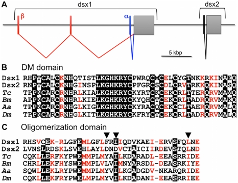
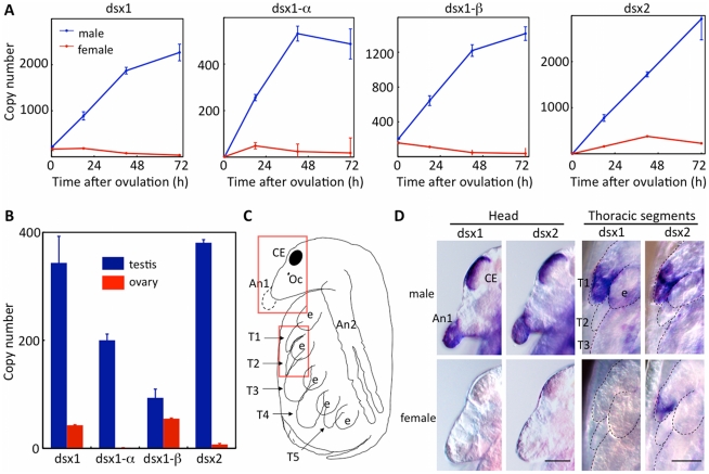
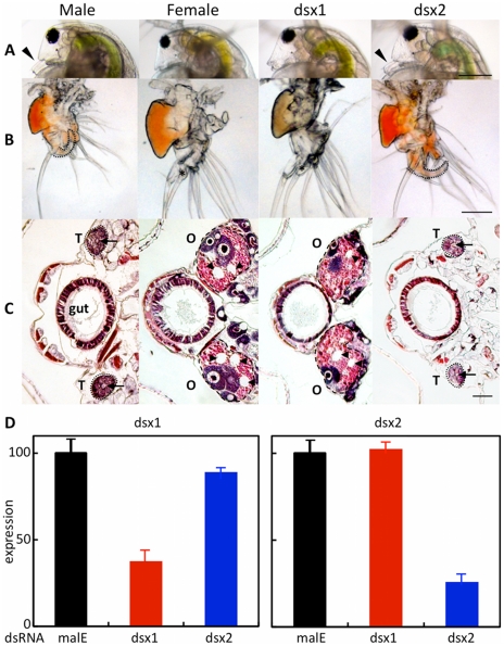
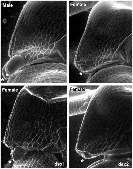
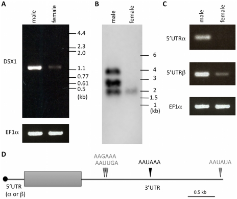
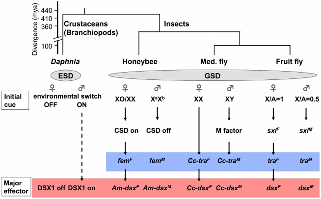
Sex-determining mechanisms are diverse among animal lineages and can be broadly divided into two major categories: genetic and environmental. In contrast to genetic sex determination (GSD), little is known about the molecular mechanisms underlying environmental sex determination (ESD). The Doublesex (Dsx) genes play an important role in controlling sexual dimorphism in genetic sex-determining organisms such as nematodes, insects, and vertebrates. Here we report the identification of two Dsx genes from Daphnia magna, a freshwater branchiopod crustacean that parthenogenetically produces males in response to environmental cues. One of these genes, designated DapmaDsx1, is responsible for the male trait development when expressed during environmental sex determination. The domain organization of DapmaDsx1 was similar to that of Dsx from insects, which are thought to be the sister group of branchiopod crustaceans. Intriguingly, the molecular basis for sexually dimorphic expression of DapmaDsx1 is different from that of insects. Rather than being regulated sex-specifically at the level of pre-mRNA splicing in the coding region, DapmaDsx1 exhibits sexually dimorphic differences in the abundance of its transcripts. During embryogenesis, expression of DapmaDsx1 was increased only in males and its transcripts were primarily detected in male-specific structures. Knock-down of DapmaDsx1 in male embryos resulted in the production of female traits including ovarian maturation, whereas ectopic expression of DapmaDsx1 in female embryos resulted in the development of male-like phenotypes. Expression patterns of another D. magna Dsx gene, DapmaDsx2, were similar to those of DapmaDsx1, but silencing and overexpression of this gene did not induce any clear phenotypic changes. These results establish DapmaDsx1 as a key regulator of the male phenotype. Our findings reveal how ESD is implemented by selective expression of a fundamental genetic component that is functionally conserved in animals using GSD. We infer that there is an ancient, previously unidentified link between genetic and environmental sex determination.


Conflict of interest statement

The authors have declared that no competing interests exist.

Figures

Figure 1
Figure 1. Daphnia Dsx genes.
(A) Genomic organization of two dsx genes in Daphnia magna. Coding exons encoding Dsx1 and Dsx2 are indicated as grey boxes, respectively. Exons comprising DSX1-α, DSX1-β and DSX2 5'UTR are indicated as blue, red and black bars. (B, C) Alignment of deduced amino acid sequences of DM domains and oligomerization domains of dsx genes. Amino acid sequences were aligned using CLUSTAL-W. Identical amino acids are highlighted in black. Similar amino acids are shown in red. Positions of non-polar amino acids important in formation of the hydrophobic interface between oligomerization domains in Drosophila Dsx protein were indicated with solid triangles . Tc, Tribolium castaneum: Bm, Bombyx mori; Aa, Aedes aegypti; Dm, Drosophila melanogaster.
Figure 2
Figure 2. Temporal and spatial dimorphic gene expression of Dsx genes during development.
(A) Male and female embryos were obtained and gene expression levels of Dsx1 and Dsx2 were determined at 18 h, 42 h and 72 h after ovulation by quantitative RT-PCR using primers corresponding to Dsx1 and Dsx2 coding sequence (CDS). Dsx1-α and DSX1-β transcripts were also quantified using primers specific to each 5'UTR. Copy numbers were estimated by quantification compared with an external standard and dividing by the number of embryos used. Bars indicate S.E.M. (B) Expression of dsx genes in adult gonads was quantified. PCR primers corresponding to Dsx1 CDS, two types of 5'UTRs of Dsx1 gene and Dsx2 CDS were used for quantitative PCR. Bars indicate S.E.M. (C) Schematic illustration of D. magna late embryo. The red boxes indicate the areas shown in panel D. (D) Whole mount in situ hybridization in late embryos using DIG-labeled probes corresponding to Dsx1 and Dsx2. The heads and the thoracic segments are magnified. Bars indicate 50 µm (heads) and 100 µm (thoracic segments). CE: Compound eye, Oc: Ocellus, An1: First antennae, An2: Second antennae, e: Epipod, T1-5: First to fifth thoracic segments.
Figure 3
Figure 3. Dimorphic development of Daphnia magna.
Eggs induced to become males were obtained from D. magna. After injection of the synthesized dsRNA, sexually dimorphic phenotypes were examined at the fifth or sixth instar except first antennae (third instar). The first two columns represent normal male and female phenotypes, respectively. The third and fourth columns represent phenotypes of individuals injected with #1-dsRNA of dsx1 and dsx2, respectively. (A): Lateral view of the head. Arrowheads indicate the first antennae. (B): First thoracic limb. Dotted line shows the outline of the stout chitinized hook. A female-type long filament corresponding to the hook is labeled with an asterisk. (C): Gonad. Daphnids were embedded in paraffin and sectioned, following by standard hematoxylin and eosin staining. Dorsal is left, ventral is right. Dotted circled lines show gonads at both sides of a gut. T and O indicate testis and ovary, respectively. Arrowheads indicate large lipid droplets lying among the eosinophilic yolk granules. Arrows indicate lumens into which the mature spermatozoa are released. (D): Gene expression profile of Dsx1 and Dsx2 in embryos injected with dsRNA of Dsx1 (left panel) and in dsRNA of Dsx2 (right panel). The MalE gene from E. coli was used as a control gene. Bars in (A), (B) and (C) indicate 200, 100, 50 µm, respectively.
Figure 4
Figure 4. Elongation of 1st antenna by the expression of Dsx1 gene.
The mRNA was injected to embryos within one hour after ovulation and observed using electron microscope after 72 h. Male and female indicate normal phenotype of each sex. Dsx1 and Dsx2 indicate mRNA of Dsx1 and Dsx2, which were injected to female eggs, respectively. Asterisk indicates first antennae. Bar indicates 100 µm.
Figure 5
Figure 5. Structure of Dsx1 mRNAs expressed in males and females.
(A) RT-PCR using oligonucleotides corresponding to 5'- and 3'-ends of Dsx1 CDS. The amplified cDNAs were resolved by agarose gel electrophoresis. (B) A northern blot probed for dsx1 mRNAs. Migration of markers with lengths indicated (kb) is shown at the right. (C) RT-PCR using oligonucleotides to amplify 5'UTR-α and -β of Dsx1 gene. The amplified cDNAs were resolved by agarose gel electrophoresis. (D) Schematic illustration of Dsx1 mRNAs with alternative isoforms due to usage of alternative promoters and polyadenylation signals. A grey box shows protein coding region; black line represents untranslated regions. Canonical and non-canonical functional polyadenylation signals identified by 3'RACE are indicated with black and grey arrowheads.
Figure 6
Figure 6. Simplified view of sex-determining pathways in the branchiopod crustacean Daphnia and insects.
An ESD pathway in Daphnia is compared with GSD pathways in insect model species, honeybee (Apis mellifera [64]), Mediterranean fruit fly (Med. fly, Ceratitis capitata, [62]) and fruit fly (Drosophila melanogaster [78]). Conserved Doublesex and Transformer homologs are indicated with red and blue boxes, respectively. Phylogenetic relationships among the four species are shown above the pathways , , . CSD, complementary sex determiner; fem, feminizer; Am, Apis mellifera: Cc, Ceratits capitata; sxl, sex lethal; mya, million years ago.

References

    1. Marin I, Baker BS. The evolutionary dynamics of sex determination. Science. 1998;281:1990–1994. - PubMed
    1. Zarkower D. Establishing sexual dimorphism: conservation amidst diversity? Nat Rev Genet. 2001;2:175–185. - PubMed
    1. Bull JJ. Sex determining mechanisms: an evolutionary perspective. Experientia. 1985;41:1285–1296. - PubMed
    1. Crews D, Bull JJ. Mode and tempo in environmental sex determination in vertebrates. Semin Cell Dev Biol. 2009;20:251–255. - PubMed
    1. Korpelainen H. Sex-Ratios and conditions required for environmental sex determination in animals. Biol Rev Camb Philos Soc. 1990;65:147–184. - PubMed

Publication types

Substances

Associated data