

α3β1 integrin promotes radiation-induced migration of meningioma cells
- PMID: 21455571
- PMCID: PMC3085848
- DOI: 10.3892/ijo.2011.987
α3β1 integrin promotes radiation-induced migration of meningioma cells
Retraction in
-
[Retracted] α3β1 integrin promotes radiation-induced migration of meningioma cells.Int J Oncol. 2021 Apr;58(4):8. doi: 10.3892/ijo.2021.5189. Epub 2021 Feb 15. Int J Oncol. 2021. PMID: 33655324 Free PMC article.
Abstract
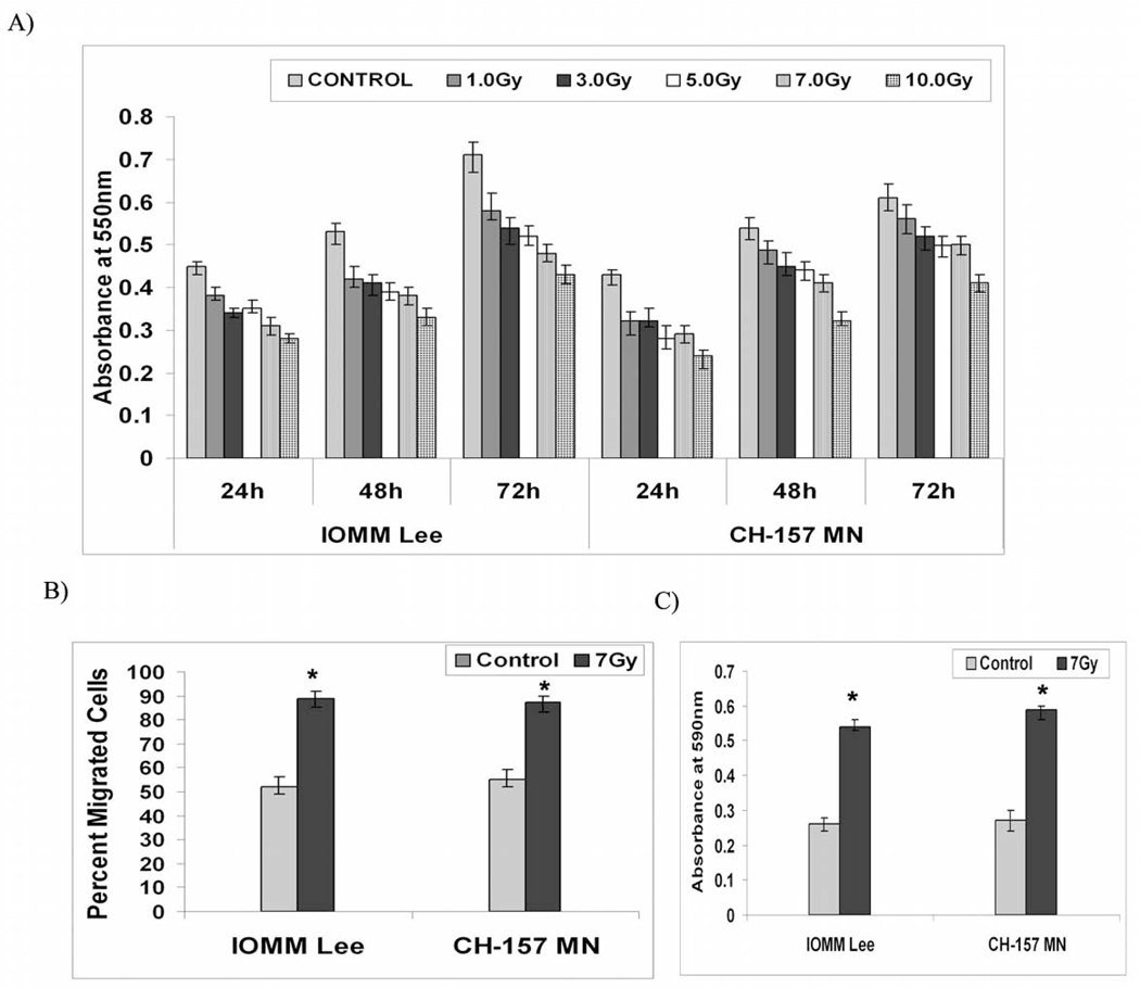
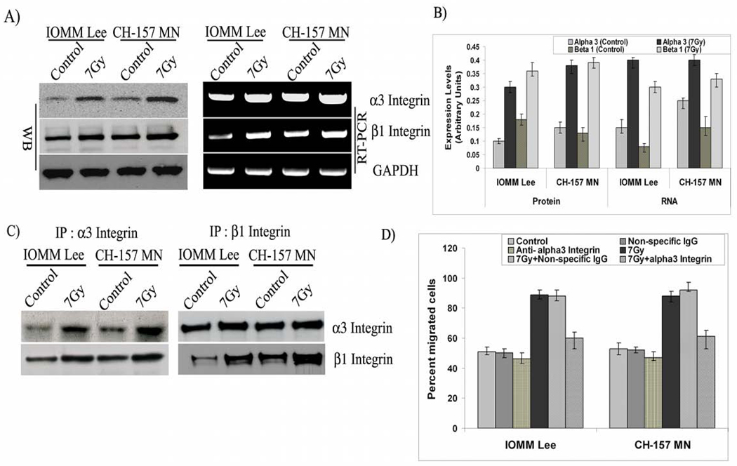
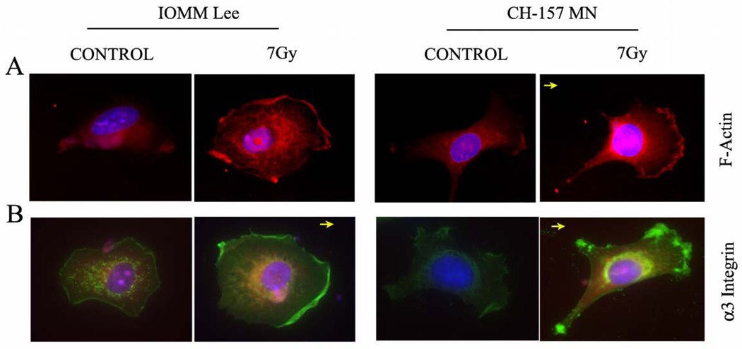
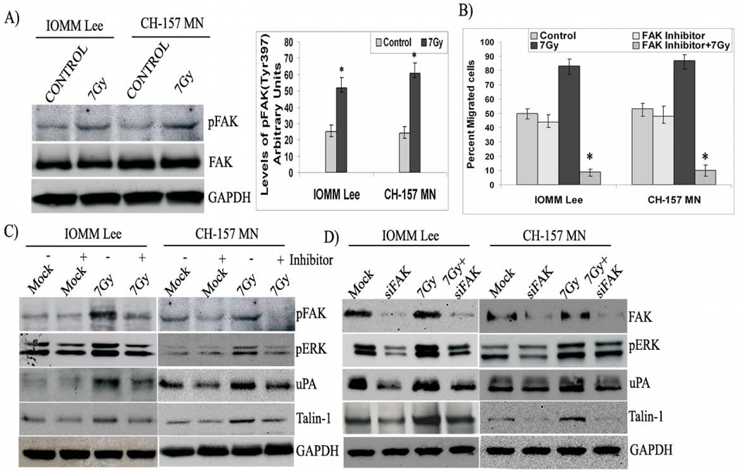
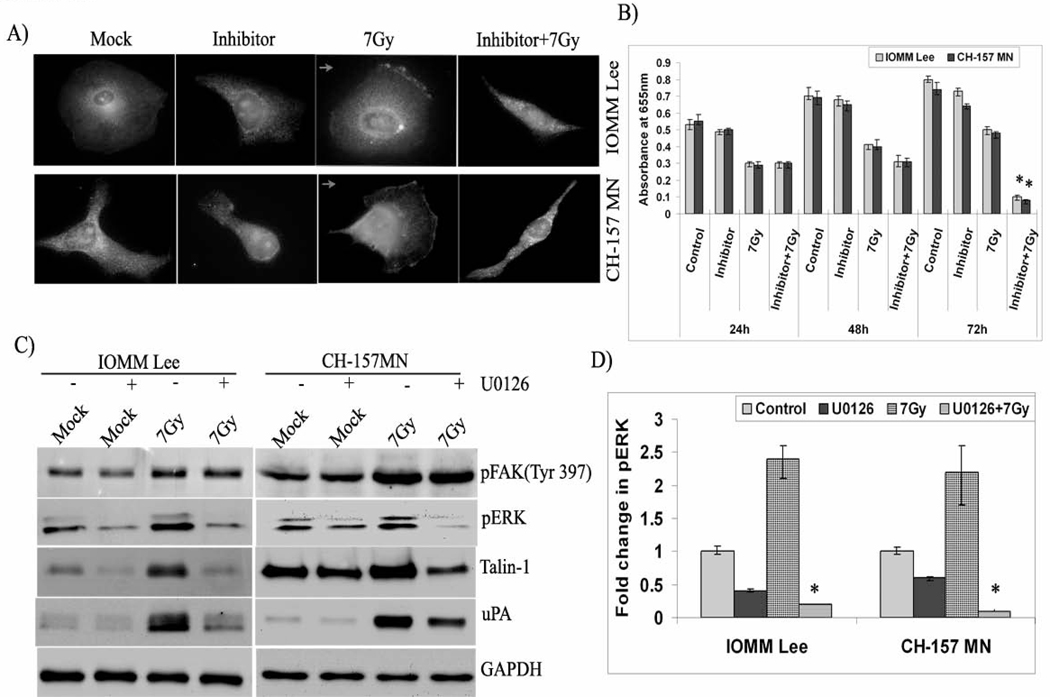
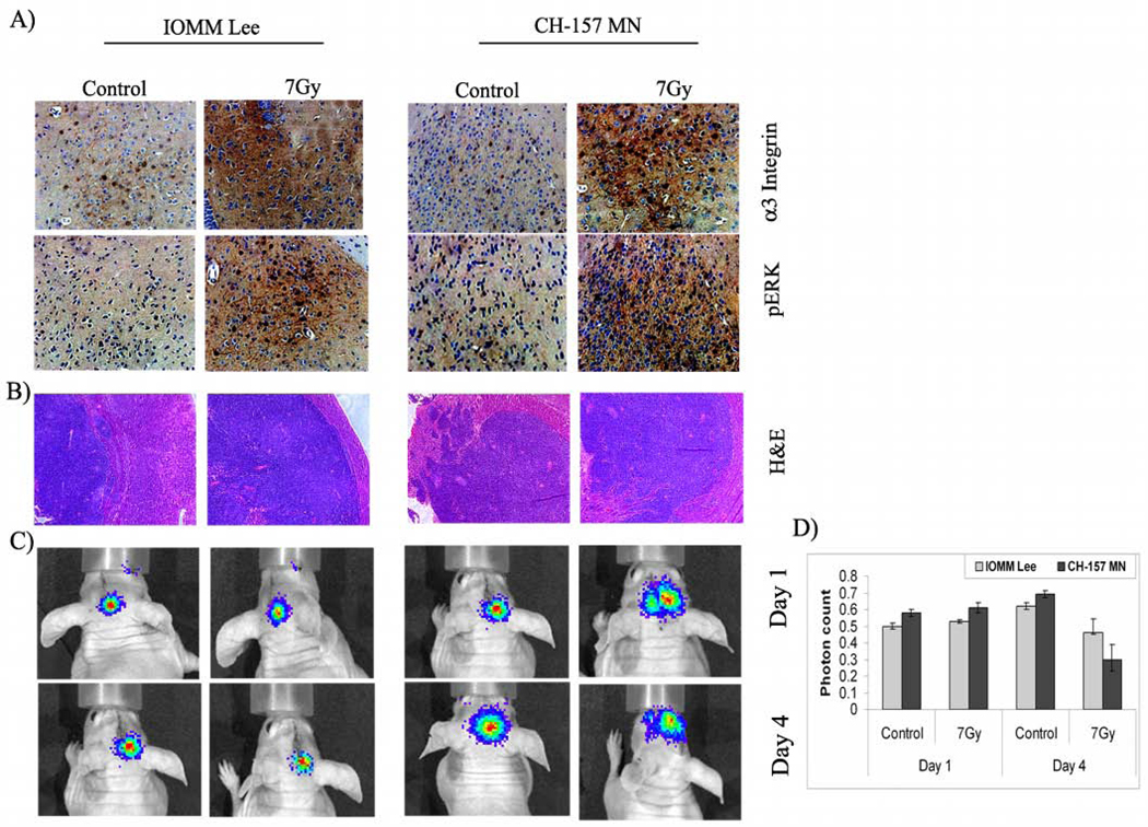
Cell motility is influenced by the microenvironment, signal transduction and cytoskeleton rearrangement. Cancer cells become resistant to these control mechanisms and gain the ability to move throughout the body and invade healthy tissues, which leads to metastatic disease. Integrins respond to context-dependent cues and promote cell migration and survival in cancer cells. In the present study, we analyzed the role of integrins in radiation-induced migration of meningioma cells. Migration and cell proliferation assays revealed that radiation treatment (7 Gy) significantly increased migration and decreased proliferation in two cell lines, IOMM-Lee and CH-157-MN. α3 and β1 integrins were overexpressed at both the protein and transcript levels after radiation treatment and a function-blocking α3β1 antibody inhibited the radiation-induced migration. Immunofluorescence studies illustrated the localization of α3 integrin and F-actin at the migration front of irradiated cells. Further, an increase in phosphorylation of FAK and ERK was observed, while both FAK phosphorylation inhibitor and FAK shRNA inhibited ERK phosphorylation and downregulated uPA and vinculin. In addition to the co-localization of FAK and ERK at the migration front, these FAK-inhibition results link the downstream effects of ERK to FAK. Correspondingly, U0126 quenched ERK phosphorylation and reduced the expression of molecules involved in migration. Furthermore, brain sections of the animals implanted with tumors followed by radiation treatment showed elevated levels of α3 integrin and active ERK. Taken together, our results show that radiation treatment enhances the migration of meningioma cells with the involvement of α3β1 integrin-mediated signaling via FAK and ERK.
Figures
Similar articles
-
Suppression of uPAR retards radiation-induced invasion and migration mediated by integrin β1/FAK signaling in medulloblastoma.PLoS One. 2010 Sep 24;5(9):e13006. doi: 10.1371/journal.pone.0013006. PLoS One. 2010. Retraction in: PLoS One. 2025 Jul 15;20(7):e0328091. doi: 10.1371/journal.pone.0328091. PMID: 20886051 Free PMC article. Retracted.
-
uPA/uPAR downregulation inhibits radiation-induced migration, invasion and angiogenesis in IOMM-Lee meningioma cells and decreases tumor growth in vivo.Int J Oncol. 2008 Nov;33(5):937-47. Int J Oncol. 2008. PMID: 18949356 Free PMC article.
-
Knockdown of cathepsin B and uPAR inhibits CD151 and α3β1 integrin-mediated cell adhesion and invasion in glioma.Mol Carcinog. 2013 Oct;52(10):777-90. doi: 10.1002/mc.21915. Epub 2012 Apr 11. Mol Carcinog. 2013. Retraction in: Mol Carcinog. 2021 Oct;60(10):717. doi: 10.1002/mc.23342. PMID: 22495828 Free PMC article. Retracted.
-
Integrin α3β1 as a breast cancer target.Expert Opin Ther Targets. 2011 Oct;15(10):1197-210. doi: 10.1517/14728222.2011.609557. Epub 2011 Aug 13. Expert Opin Ther Targets. 2011. PMID: 21838596 Free PMC article. Review.
-
Growth control by intracellular tension and extracellular stiffness.Trends Cell Biol. 2008 Jul;18(7):347-52. doi: 10.1016/j.tcb.2008.05.002. Epub 2008 May 29. Trends Cell Biol. 2008. PMID: 18514521 Free PMC article. Review.
Cited by
-
X-radiation enhances the collagen type I strap formation and migration potentials of colon cancer cells.Oncotarget. 2016 Nov 1;7(44):71390-71399. doi: 10.18632/oncotarget.12111. Oncotarget. 2016. PMID: 27655687 Free PMC article.
-
In vitro radiotherapy and chemotherapy alter migration of brain cancer cells before cell death.Biochem Biophys Rep. 2021 Jul 13;27:101071. doi: 10.1016/j.bbrep.2021.101071. eCollection 2021 Sep. Biochem Biophys Rep. 2021. PMID: 34286111 Free PMC article.
-
A Monoclonal Antibody Against β1 Integrin Inhibits Proliferation and Increases Survival in an Orthotopic Model of High-Grade Meningioma.Target Oncol. 2019 Aug;14(4):479-489. doi: 10.1007/s11523-019-00654-4. Target Oncol. 2019. PMID: 31301014
-
Lung cancer cells that survive ionizing radiation show increased integrin α2β1- and EGFR-dependent invasiveness.PLoS One. 2013 Aug 8;8(8):e70905. doi: 10.1371/journal.pone.0070905. eCollection 2013. PLoS One. 2013. PMID: 23951036 Free PMC article.
-
MMP-9 silencing regulates hTERT expression via β1 integrin-mediated FAK signaling and induces senescence in glioma xenograft cells.Cell Signal. 2011 Dec;23(12):2065-75. doi: 10.1016/j.cellsig.2011.08.001. Epub 2011 Aug 9. Cell Signal. 2011. PMID: 21855630 Free PMC article.
References
-
- Ridley AJ, Schwartz MA, Burridge K, Firtel RA, Ginsberg MH, Borisy G, Parsons JT, Horwitz AR. Cell migration: integrating signals from front to back. Science. 2003;302:1704–1709. - PubMed
-
- Cukierman E, Pankov R, Stevens DR, Yamada KM. Taking cell-matrix adhesions to the third dimension. Science. 2001;294:1708–1712. - PubMed
-
- Hood JD, Cheresh DA. Role of integrins in cell invasion and migration. Nat Rev Cancer. 2002;2:91–100. - PubMed
-
- Schmid RS, Shelton S, Stanco A, Yokota Y, Kreidberg JA, Anton ES. alpha3beta1 integrin modulates neuronal migration and placement during early stages of cerebral cortical development. Development. 2004;131:6023–6031. - PubMed
-
- Sordat I, Decraene C, Silvestre T, Petermann O, Auffray C, Pietu G, Sordat B. Complementary DNA arrays identify CD63 tetraspanin and alpha3 integrin chain as differentially expressed in low and high metastatic human colon carcinoma cells. Lab Invest. 2002;82:1715–1724. - PubMed
Publication types
MeSH terms
Substances
Grants and funding
LinkOut - more resources
Full Text Sources
Miscellaneous
