. 2011 Aug 1;519(11):2175-92.
doi: 10.1002/cne.22623.

Morphologic integration of hilar ectopic granule cells into dentate gyrus circuitry in the pilocarpine model of temporal lobe epilepsy

Affiliations

Morphologic integration of hilar ectopic granule cells into dentate gyrus circuitry in the pilocarpine model of temporal lobe epilepsy

Michael C Cameron et al. J Comp Neurol. .

Abstract

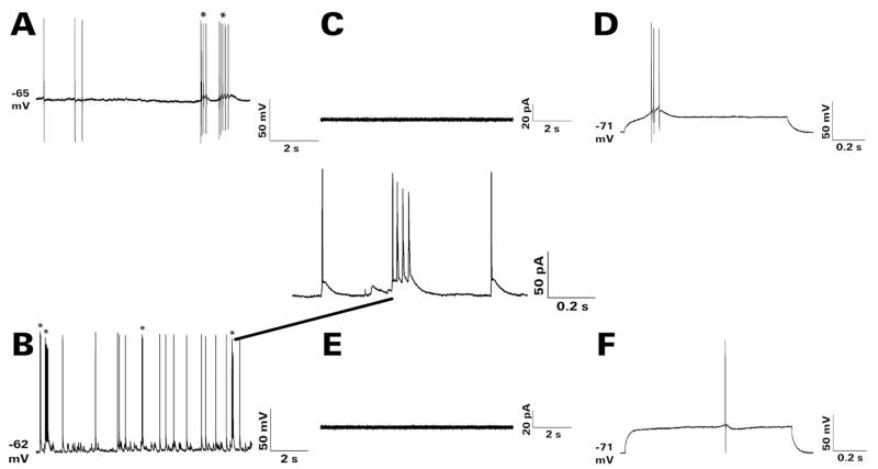
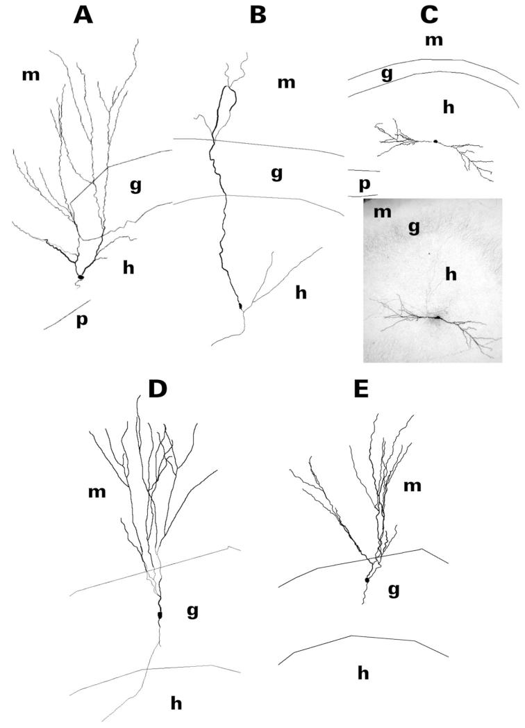
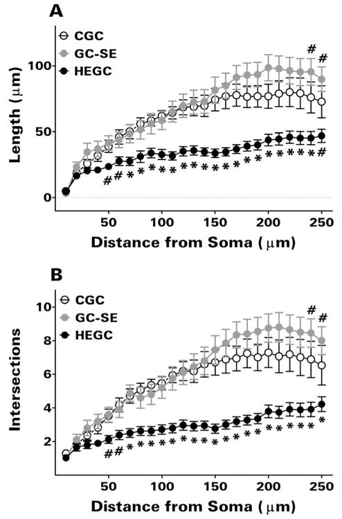
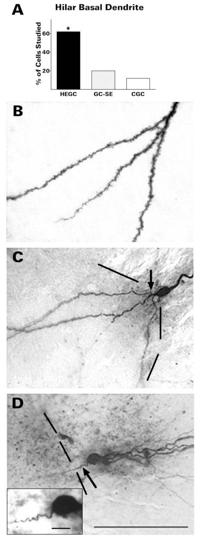
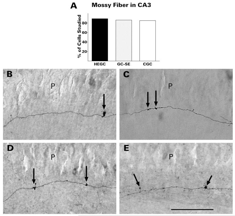
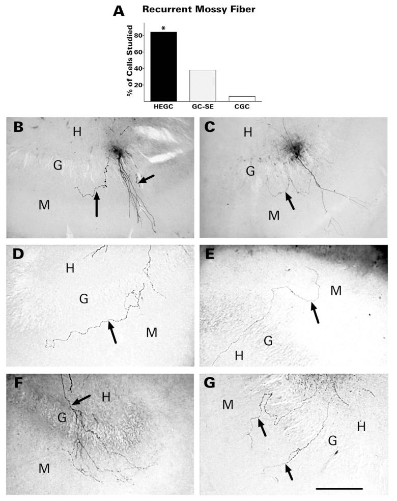
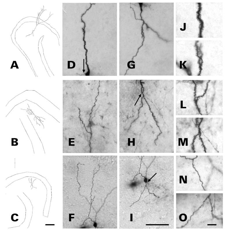
After pilocarpine-induced status epilepticus, many granule cells born into the postseizure environment migrate aberrantly into the dentate hilus. Hilar ectopic granule cells (HEGCs) are hyperexcitable and may therefore increase circuit excitability. This study determined the distribution of their axons and dendrites. HEGCs and normotopic granule cells were filled with biocytin during whole-cell patch clamp recording in hippocampal slices from pilocarpine-treated rats. The apical dendrite of 86% of the biocytin-labeled HEGCs extended to the outer edge of the dentate molecular layer. The total length and branching of HEGC apical dendrites that penetrated the molecular layer were significantly reduced compared with apical dendrites of normotopic granule cells. HEGCs were much more likely to have a hilar basal dendrite than normotopic granule cells. They were about as likely as normotopic granule cells to project to CA3 pyramidal cells within the slice, but were much more likely to send at least one recurrent mossy fiber into the molecular layer. HEGCs with burst capability had less well-branched apical dendrites than nonbursting HEGCs, their dendrites were more likely to be confined to the hilus, and some exhibited dendritic features similar to those of immature granule cells. HEGCs thus have many paths along which to receive synchronized activity from normotopic granule cells and to transmit their own hyperactivity to both normotopic granule cells and CA3 pyramidal cells. They may therefore contribute to the highly interconnected granule cell hubs that have been proposed as crucial to development of a hyperexcitable, potentially seizure-prone circuit.


Figures

Figure 1
Figure 1
Spontaneous and depolarization-evoked bursting of representative HEGCs. The recordings shown were obtained at 22-24 ° during superfusion at ≈3 mL/min with artificial cerebrospinal fluid that contained 122 mM NaCl, 25 mM NaHCO3, 3.1 mM KCl, 1.8 mM CaCl2, 1.2 mM MgSO4, 0.4 mM KH2PO4, and 10 mM D-glucose, equlibrated with 95% O2 / 5% CO2. A cellular burst was defined as two or more action potentials superimposed on a single depolarizing wave. No such events were detected in recordings from any GC-SE or CGC made under these conditions. Cellular bursts are indicated by asterisks (*) in panels A, B. A: Gramicidin perforated patch recording in current clamp mode. Spontaneous action potentials and cellular bursts were recorded at the resting membrane potential of −65 mV. B: Whole-cell patch clamp recording in current clamp mode obtained soon after break-in. Spontaneous action potentials and bursts were recorded at the resting membrane potential of −62 mV. The expanded trace shows the fourth spontaneous burst in greater detail. C,D: Depolarization-evoked burst recorded during whole-cell patch clamp recording in current clamp mode. C: Membrane currents were monitored for 5 minutes during cell-attached recording in voltage clamp mode at a holding potential of −70 mV. No currents associated with spontaneous bursting were recorded from this cell. D: After achieving whole cell access, membrane potential was monitored during successive injections of depolarizing current from −0.5 to +1.9 nA for 1 second each. The cell’s minimal response was a burst of action potentials first recorded during depolarization to −56 mV from a resting membrane potential of −71 mV. E,F: Lack of depolarization-evoked bursting in another recorded HEGC. E: Again, no currents associated with spontaneous bursting were observed by cell-attached recording. In addition, no spontaneous bursts were recorded at the resting membrane potential upon achieving whole cell access. F: Depolarization of this cell to −56 mV from a resting membrane potential of −71 mV evoked no response. Its minimal response was a single action potential first recorded during depolarization to −43 mV. In these respects, this nonbursting HEGC resembled closely all recorded GC-SEs and CGCs. CGC, normotopic granule cell from control rat; GC-SE, normotopic granule cell from rat that had experienced status epilepticus; HEGC, hilar ectopic granule cell from rat that had experienced status epilepticus.
Figure 2
Figure 2
Reconstruction of dentate granule cell somatodendritic morphology after filling the cell with biocytin during whole-cell patch clamp recording in hippocampal slices. After visualization of biocytin with avidin/horseradish peroxidase/diaminobenzidine, cells were reconstructed from serial sections with use of Neurolucida. Granule cells shown are representative of those cells having a hilar basal dendrite. HEGCs shown (A–C) were chosen to illustrate differences in the relative sizes of the apical and basal dendritic trees: predominantly apical (A), mainly apical (B), and apical and basal about equal (C). The apical dendrite of HEGCs could reach the outer edge of the dentate molecular layer (A,B) or be confined to the dentate hilus (C; the dendrite pointed to the left was considered the apical dendrite). Note the relatively sparse branching of HEGC apical dendrites compared to those of GE-SEs (D) and CGCs (E) and the relatively exuberant branching of the distal apical dendrite(s) of GC-SEs compared to that of CGCs. A photomicrograph of the HEGC in panel C is included for comparison with the reconstruction. CGC, normotopic granule cell from control rat; g, granule cell body layer; GC-SE, normotopic granule cell from rat that had experienced status epilepticus; h, hilus of the dentate gyrus; HEGC, hilar ectopic granule cell from rat that had experienced status epilepticus; m, molecular layer of the dentate gyrus; p, pyramidal cell body layer of area CA3c.
Figure 3
Figure 3
Scholl analysis revealed significantly less apical dendritic length (A) and branching (B) in HEGCs than in normotopic granule cells beginning at 50 μm from the soma. This analysis also confirmed the greater length and branching of the distal apical dendrite in GC-SEs than in CGCs. Only HEGCs whose apical dendrite(s) penetrated through the dentate molecular layer were included. Values are means ± SEM for 34 HEGCs, 20 GC-SEs, and 17 CGCs. #P <0.05 or *P < 0.01 compared with the other two groups by Newman-Keuls test after two-way ANOVA (granule cell group × dendritic segment) yielded P <0.001 for granule cell group, dendritic segment, and the interaction between these two variables. CGC, normotopic granule cells from control rats; GC-SE, normotopic granule cells from rats that had experienced status epilepticus; HEGC, hilar ectopic granule cells from rats that had experienced status epilepticus.
Figure 4
Figure 4
HEGCs were much more likely to have a hilar basal dendrite than normotopic dentate granule cells (A). The hilar basal dendrite of HEGCs (B) was usually heavily invested with spines and branched either close to the soma, more distally, or both. The spiny hilar basal dendrite of GC-SEs (C) branched once close to the soma (arrow) or not at all. In contrast, the few basal dendrites observed on CGCs (D) were relatively short and unbranched, without visible spines (arrow). The inset in panel D illustrates the morphology of the CGC hilar basal dendrite at higher magnification. Dashed lines indicate the border between the granule cell body layer and hilus. Quantitative data were derived from microscopic observation of 79 HEGCs, 54 GC-SEs, and 40 CGCs. *P <0.001 compared with the other two groups by χ-square test. CGC, normotopic granule cells from control rats; GC-SE, normotopic granule cells from rats that had experienced status epilepticus; HEGC, hilar ectopic granule cells from rats that had experienced status epilepticus. Scale bars 100 μm; inset, 10 μm.
Figure 5
Figure 5
HEGCs were about as likely as normotopic dentate granule cells to project to area CA3 neurons within the slice (A). Mossy fibers of HEGCs (D,E), like those of granule cells from control rats (B) and GC-SEs (C), had widely spaced giant boutons in stratum lucidum from which filopodia emerged (arrows). Quantitative data were derived from microscopic observation of 55 HEGCs, 32 GC-SEs, and 33 CGCs. CGC, normotopic granule cells from control rats; GC-SE, normotopic granule cells from rats that had experienced status epilepticus; HEGC, hilar ectopic granule cells from rats that had experienced status epilepticus; P, pyramidal cell body layer. Scale bar = 200 μm.
Figure 6
Figure 6
HEGCs were much more likely to send one or more recurrent mossy fibers (arrows) into the dentate molecular layer than normotopic granule cells (A). B,C: Recurrent mossy fibers of GC-SEs. In panel B the labeled cell has two visible recurrent mossy fibers in the molecular layer, one of which courses through the inner third of the molecular layer parallel to the granule cell body layer. In panel C, one recurrent fiber branches in the granule cell body layer and the two branches course in opposite directions through the inner molecular layer. D-G: Recurrent mossy fibers of HEGCs. D: A single recurrent mossy fiber courses through the inner third of the molecular layer parallel to the granule cell body layer. E: The fiber runs for a short distance through the inner third of the molecular layer then extends through the outer part of the layer. F: A recurrent mossy fiber branches extensively in the granular and molecular layers. G: Two separate fibers emerge from the hilus and cross the granule cell body layer. Quantitative data were derived from microscopic observation of 55 HEGCs, 32 GC-SEs, and 33 CGCs. *P <0.001 compared to the other two groups by χ-square test. CGC, normotopic granule cells from control rats; G, granule cell body layer; GC-SE, normotopic granule cells from rats that had experienced status epilepticus; H, hilus of the dentate gyrus; HEGC, hilar ectopic granule cells from rats that had experienced status epilepticus; M, molecular layer of the dentate gyrus. Scale bar = 200 μm.
Figure 7
Figure 7
Dendritic morphology of three bursting HEGCs. Cells were filled with biocytin during whole-cell patch clamp recording and cell morphology was reconstructed with use of Neurolucida. Micrographs of the apical (D–F) and basal (G-I) dendrites of the three representative HEGCs are shown to the right of each reconstruction (A-C). Arrows indicate the origin of the mossy fiber. The micrographs to the far right of each reconstruction illustrate the morphology of apical (J,L,N) and basal (K,M,O) dendrites at higher magnification. Dendrites of HEGC A exhibit morphology typical of both bursting and nonbursting HEGCs. The apical dendritic branches reach the outer edge of the molecular layer and both apical and hilar basal dendrites appear fully formed and heavily invested with spines. Dendrites of HEGC B are confined to the hilus, but dendritic structure appears normal otherwise. The varicose dendrites of HEGC C are confined to the hilus, are unusually short, and lack mature spines. Scale bars = 200 μm left; 70 μm center; 10 μm right.

Similar articles

Cited by

References

    1. Acsády L, Kamondi A, Sik A, Freund T, Buzsáki G. GABAergic cells are the major postsynaptic targets of mossy fibers in the rat hippocampus. J Neurosci. 1998;18:3386–3403. - PMC - PubMed
    1. Ambrogini P, Lattanzi D, Ciuffoli S, Agostini D, Bertini L, Stocchi V, Santi S, Cuppini R. Morpho-functional characterization of neuronal cells at different stages of maturation in granule cell layer of adult dentate gyrus. Brain Res. 2004;1017:21–31. - PubMed
    1. Bonde S, Ekdahl CT, Lindvall O. Long-term neuronal replacement in adult rat hippocampus after status epilepticus despite chronic inflammation. Eur J Neurosci. 2006;23:965–974. - PubMed
    1. Bough KJ, Mott DD, Dingledine RJ. Medial perforant path inhibition mediated by mGluR7 is reduced after status epilepticus. J Neurophysiol. 2004;92:1549–1557. - PubMed
    1. Buckmaster PS, Dudek FE. In vivo intracellular analysis of granule cell axon reorganization in epileptic rats. J Neurophysiol. 1999;81:712–721. - PubMed

Publication types

MeSH terms