. 2011 Aug 15;519(12):2434-74.
doi: 10.1002/cne.22635.

Vasopressin innervation of the mouse (Mus musculus) brain and spinal cord

Affiliations

Vasopressin innervation of the mouse (Mus musculus) brain and spinal cord

Benjamin D Rood et al. J Comp Neurol. .

Abstract

The neuropeptide vasopressin (AVP) has been implicated in the regulation of numerous physiological and behavioral processes. Although mice have become an important model for studying this regulation, there is no comprehensive description of AVP distribution in the mouse brain and spinal cord. With C57BL/6 mice, we used immunohistochemistry to corroborate the location of AVP-containing cells and to define the location of AVP-containing fibers throughout the mouse central nervous system. We describe AVP-immunoreactive (-ir) fibers in midbrain, hindbrain, and spinal cord areas, which have not previously been reported in mice, including innervation of the ventral tegmental area, dorsal and median raphe, lateral and medial parabrachial, solitary, ventrolateral periaqueductal gray, and interfascicular nuclei. We also provide a detailed description of AVP-ir innervation in heterogenous regions such as the amygdala, bed nucleus of the stria terminalis, and ventral forebrain. In general, our results suggest that, compared with other species, the mouse has a particularly robust and widespread distribution of AVP-ir fibers, which, as in other species, originates from a number of different cell groups in the telencephalon and diencephalon. Our data also highlight the robust nature of AVP innervation in specific regulatory nuclei, such as the ventral tegmental area and dorsal raphe nucleus among others, that are implicated in the regulation of many behaviors.


Figures

Figure 1
Figure 1
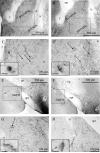
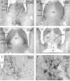
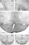
BNST and amygdala. A: Bregma –0.10. Small, lightly staining AVP-ir cells are found above the fibers of the stria terminalis in the medial BSTMPM. B: Bregma –0.22. AVP-ir cells are also found in the BSTMPL slightly caudal and ventral to those in the BSTMPM. C: Magnification of the BSTMPM from boxed area in A. D: Magnification of the BSTMPL from boxed area in B. E: Bregma –1.34. AVP-ir cells are found at the dorsolateral edge of the rostral MePD. F: Bregma –1.46. AVP-ir cells are found in the caudal MePD as well. G: Magnification of the rostral MePD from boxed area in E. H: Magnification of the caudal MePD from boxed area in F. In C,D,G,H, arrows indicate AVP-ir cells, and insets depict individual cells at high magnification. Scale bars = 500 μm in A,B,E,F; 100 μm in C,D,G,H; 10 μm in insets.
Figure 2
Figure 2
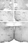
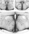
PVN and SON. A: Bregma –0.46. AVP-ir neurons in the PaAP. B: Bregma –0.70. Magnocellular neurons are located close to the third ventricle in the PaMM and PaV. C: Bregma –0.82. Magnocellular neurons in the PaLM, and parvocellular neurons in the PaMP medially near the ventricle. D: Magnocellular neurons in the PaLM persist as other cell groups end caudally. E: Bregma –0.58. AVP-ir cells in the episupraoptic nucleus just dorsal to the rostral tip of the optic tract (opt). F: Bregma –0.82. Rostral pole of extremely dense cluster of AVP-ir cells in the SON dorsal and lateral to the optic tract. G: Bregma –0.94. Continuation of dense cluster of AVP-ir cells dosal and lateral to the lateral edge of the optic tract, plus AVP-ir cells along the ventral brain surface closer to the midline. H: Bregma –1.06. A few scattered cells lateral to the optic tract at the caudal edge of the SON. Scale bar = 200 μm.
Figure 3
Figure 3
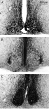
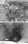
SCN. A: Bregma –0.34. The rostral SCN contains a dense cluster of small, darkly staining cells. B: Bregma –0.58. In the middle SCN, AVP-ir cells in the SCN form a shell around an AVP-ir cell-free core. C: Bregma –0.82. The caudal SCN also has a dense cluster of AVP-ir cells. Outlines illustrate the extent of cells in the SCN. Scale bar = 100 μm.
Figure 4
Figure 4
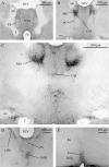
Accessory nuclei and other hypothalamic nuclei. Darkly staining AVP-ir neurons in the rostral anterior hypothalamus (AHA; bregma –0.46; A), perifornical region (bregma –0.70; B), peduncular lateral hypothalamus (PLH; bregma –1.22; C), “mouse accessory nucleus” (Castel and Morris, 1988) in the striohypothalamic region (StHy; bregma –0.22; D), nucleus circularis (NC; bregma –0.46; E), rostral periventricular area (Pe; bregma –0.10; F), ventral lateral preoptic area (VLPO; bregma 0.02; G), and retrochiasmatic supraoptic nucleus at the level of the tuberal lateral hypothalamus (SOR; bregma –1.46; H). In all pictures, arrows indicate representative cells within the picture. Scale bars = 100 μm in A,D,G; 200 μm in B,C,H; 50 μm in E,F.
Figure 5
Figure 5
Fiber size. A: Bregma 0.38. Small AVP-ir fibers in the LSV with punctate staining. B: Bregma –4.72. Medium-sized fibers in the VLPAG with occasional terminal boutons with visible stalks (see circled structure in B). In each figure, arrows indicate representative fibers. Scale bars = 100 μm in A,B; 25 μm in insets.
Figure 6
Figure 6
Cortex and hippocampus. A–C: Sparse medium-sized AVP-ir fibers in the the PrL (bregma 1.98; A), insular cortex (bregma – 0.10; B), and Cg2 (bregma 1.10; C). D: Bregma –2.54. Small fibers are seen in more caudal regions of the cortex, especially the DLEnt. E: Bregma –2.92. Darkfield image of CA1 region of the ventral hippocampus. F: Brightfield image showing small fibers from boxed area in E. G: Bregma –3.28. Darkfield image of CA1 region of the ventral hippocampus. H: Brightfield image showing medium-sized fibers from boxed area in G. Scale bars = 200 μm in A,C,F; 100 μm in B,H; 500 μm in D,E,G.
Figure 7
Figure 7
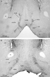
Basal ganglia and ventral forebrain. A: Bregma 0.98. Small AVP-ir fibers in the VP and the ventral septal area (VSA) between the AcbSh and VDB in the rostral forebrain. Medium-sized fibers are found in the AcbC. B: Bregma 0.6. Small fibers in the MPA, VP, SI, and VSA. AVP-ir fibers avoid the VDB, ICjM, Tu, and AcbSh. Scale bars = 200 μm.
Figure 8
Figure 8
Septal complex. A: Bregma 0.86. Sparse medium-sized AVP-ir fibers in the rostral LS. B: Bregma 0.62. Small fibers in the LSV and LSI. C: Bregma 0.38. Very dense small fiber innervation of the caudal part of the LSV and LSI. D: Bregma –0.1. Isolated small fibers in the SFi and medium-sized fibers in the SFO. E: Magnification of the LSV from B. F: High magnification of boxed area in E. The arrow indentifies a pericellular basket. Scale bars = 500 μm in A–D; 200 μm in E; 100 μm in F.
Figure 9
Figure 9
Bed nucleus of the stria terminalis. A: Bregma 0.50. Small and medium-sized AVP-ir fibers in the BSTMA and medium-sized fibers in the BSTMV. B: Bregma 0.02. Small fibers in the BSTLV and medium-sized fibers in the BSTMV. C: Bregma –0.10. Two distinct clusters of small fibers and a number of medium-sized fibers in the BSTMPM, small fibers in the BSTLV, and medium-sized fibers in the BSTMV. D: Bregma –0.34. BSTMPL with BNST cells in the BSTMPL and small fibers, which can be seen entering the stria terminalis (st). E: Bregma –0.46. Dense small fibers ventral to stria medullaris (sm), but less dense in the rest of the BNST. Scale bars = 100 μm.
Figure 10
Figure 10
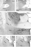
Amygdala. A: Bregma –0.58. Diffuse small fibers in the AA and EA (see footnote 1). B: Magnification from boxed area in A showing moderate numbers of fibers in the EA and a denser plexus in the neighboring PLH. C: Bregma –0.7. Small fibers in the EA, AA, and ACo but not in the LOT. D: Magnification from boxed area in C. E: Bregma –1.3. Medium-sized fibers in CeA. F: Bregma –1.58. A dense small fiber plexus is found in the MePD. G: Bregma –2.18. Small fibers in the caudal BLP just ventral to the LA. H: Magnification from boxed area in G showing fibers in the BLP. Scale bars = 500 μm in A,C,G; 200 μm in B,D,H; 100 μm in E,F.
Figure 11
Figure 11
Preoptic area. A: Bregma 0.38. Small AVP-ir fibers in the MPA and medium-sized fibers in the VOLT and MnPO. B: Bregma 0.26. MnPO at higher magnification. Medium-sized fibers are oriented vertically in the plane of section. C: Bregma 0.14. Dense small fiber plexus in the PS ventral to the BSTMV. D: Magnification of boxed area in E showing scattered small and medium-sized fibers in the MPOM, and many small fibers and some medium-sized fibers in AVPe. E: Bregma –0.10. Sparse small and medium-sized fibers in the caudal LPO and StHy. Scale bars = 500 μm in A,E; 200 μm in B; 100 μm in C,D.
Figure 12
Figure 12
Anterior hypothalamus. A: Bregma –0.58. Small fibers in the AHA ventral to the fornix and dorsal to the SCN adjacent to the third ventricle and medium-sized fibers stretching between the PVN and SON through ventral parts of the AHA. B: Bregma –0.82. Small fibers are found medially in the SCN, SPa, and PVN. Medium-sized fibers dominate the AHC and numerous medium-sized fibers pass over the fornix into the PLH. C: Bregma –0.94. Caudally, fewer medium-sized fibers pass through the AHC, but many still pass into the PLH. Large amounts of small fibers are still found along the third ventricle. The dotted line indicates the approximate location of the sagittal section shown in Figure 13. Scale bars = 500 μm.
Figure 13
Figure 13
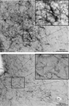
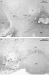
Sagittal section of hypothalamus immediately lateral to the third ventricle (see dotted line in Fig. 12 for approximate location). A: Large, medium-sized, and small fibers can be seen among the many PVN neurons adjacent to the third ventricle (large, medium-sized, and small vertical arrows indicate fibers of each type). The last white arrow identifies a lighter region that corresponds to the third ventricle. B: In the same sagittal section, a dense cloud of small fibers is seen in and around the SCN. Fibers can be seen passing in all direction, with the largest groups passing dorsally, dorsocaudally, and rostrodorsally. Scale bars = 200 μm.
Figure 14
Figure 14
Dorsomedial hypothalamus. A: Bregma –1.46. Small and medium-sized fibers in the DM and Arc, medium-sized fibers in the TuLH, and a near absence of fibers in the VMH. B: Bregma –1.82. Small and medium-sized fibers in the DM. C: Magnification of DM from boxed area in A. Note small punctate staining closer to 3V and numerous medium-sized fibers (arrows). D: Magnification of the ME from B. Note dense staining in dorsal ME (inner zone) and less dense staining in the ventral ME (outer zone). Scale bars = 500 μm in A,B; 100 μm in C,D.
Figure 15
Figure 15
Lateral hypothalamus and mammillary bodies. A: Bregma –0.82. Small fibers among medium-sized fibers passing through the PLH. B: Bregma –1.2:. Fewer small fibers are seen in the caudal PLH, but a dense array of medium-sized and large fibers is observed. C: Bregma –2.46. Small fibers in the PeFLH and DTM around the fornix (f). Scattered small and medium-sized fibers in the PMV, PH, and PSTh. D: Bregma –2.54. Dense plexus of small fibers just ventral to the MB, small and medium-sized fibers in RMM, scattered small fibers in the PMV, and absence of fibers in MB. E: Bregma –2.80. Dense plexus of small fibers in the VTM and scattered small and medium-sized fibers in the RMM. Scale bars = 100 μm in A; 200 μm in B; 500 μm in C–E.
Figure 16
Figure 16
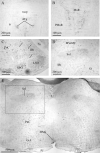
Thalamus. A: Bregma –0.34. Small fibers in the PVA and periphery of the PT, Arrow points at a dense cluster of fibers dorsal to the PT. B: Bregma –0.58. Dense plexus of small fibers in the MD and additional fibers in the CM and PC surrounding the PT. C: Bregma –0.94. Dense plexus of small fibers in the MD and additional small fibers in the PV, CM, and Rh. Medium-sized fibers in the PV and Rh. D: Bregma –1.22. Small fibers in the LHb and IAD and in the PV just ventral to the dorsal aspect of the third ventricle. E: Bregma –1.2. Small fibers in the Xi just dorsal to the third ventricle. F: Magnification of MD from B illustrates density of fibers in this region. G: Bregma –1.58. Dense cluster of small fibers in the LHb, with light innervation of the PV and IMD. H: A sagittal section (lateral 0.48 mm) shows the pathway of AVP-ir fibers arching dorsally just ventral to the stria medullaris, with dense fiber plexuses in the LHb and MD. I: Bregma –0.58. Small fibers in the ZI. J: Bregma –1.8. Scattered small fibers in the caudal extent of the LHb, with primarily medium-sized fibers in the PV. Scale bars = 500 μm in A–C,G,H; 200 μm in D,E,I,J; 100 μm in F.
Figure 16
Figure 16
Thalamus. A: Bregma –0.34. Small fibers in the PVA and periphery of the PT, Arrow points at a dense cluster of fibers dorsal to the PT. B: Bregma –0.58. Dense plexus of small fibers in the MD and additional fibers in the CM and PC surrounding the PT. C: Bregma –0.94. Dense plexus of small fibers in the MD and additional small fibers in the PV, CM, and Rh. Medium-sized fibers in the PV and Rh. D: Bregma –1.22. Small fibers in the LHb and IAD and in the PV just ventral to the dorsal aspect of the third ventricle. E: Bregma –1.2. Small fibers in the Xi just dorsal to the third ventricle. F: Magnification of MD from B illustrates density of fibers in this region. G: Bregma –1.58. Dense cluster of small fibers in the LHb, with light innervation of the PV and IMD. H: A sagittal section (lateral 0.48 mm) shows the pathway of AVP-ir fibers arching dorsally just ventral to the stria medullaris, with dense fiber plexuses in the LHb and MD. I: Bregma –0.58. Small fibers in the ZI. J: Bregma –1.8. Scattered small fibers in the caudal extent of the LHb, with primarily medium-sized fibers in the PV. Scale bars = 500 μm in A–C,G,H; 200 μm in D,E,I,J; 100 μm in F.
Figure 17
Figure 17
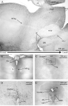
Tegmentum. A: Bregma –3.08. Small and medium-sized fibers in the VTA, IF and ventromedial pole of the SNR. B: Bregma – 3.52. Small fibers surrounding the IPR in the IF and PIF. C: Bregma –4.04. Dense plexus of medium-sized fibers and isolated small fibers in the caudal tegmentum in the mRt and RRF just dorsal to the SNR. D: Bregma –4.04. Predominantly small fibers n the CLi between the superior cerebellar peduncles (scp). E: Bregma –4.04. Moderate numbers of small fibers in the caudal pole of the IP. Scale bars = 200 μm in A–D; 100 μm in E.
Figure 17
Figure 17
Tegmentum. A: Bregma –3.08. Small and medium-sized fibers in the VTA, IF and ventromedial pole of the SNR. B: Bregma – 3.52. Small fibers surrounding the IPR in the IF and PIF. C: Bregma –4.04. Dense plexus of medium-sized fibers and isolated small fibers in the caudal tegmentum in the mRt and RRF just dorsal to the SNR. D: Bregma –4.04. Predominantly small fibers n the CLi between the superior cerebellar peduncles (scp). E: Bregma –4.04. Moderate numbers of small fibers in the caudal pole of the IP. Scale bars = 200 μm in A–D; 100 μm in E.
Figure 18
Figure 18
Periaqueductal gray and dorsal raphe nuclei. A: Sagittal section near the midline showing AVP-ir pathways travelling dorsally toward the PAG through the rostral midbrain (left arrow) and caudally through the IF and CLi (right arrow) toward the DR. B: Bregma –2.30. Small and medium-sized fibers ascend dorsally between the right and left fasciculus retroflexus (fr) immediately rostral to PAG. C: Bregma –3.52. Plexus of small fibers in the DMPAG. D: Magnification of small fibers in the DMPAG from boxed area in C. E: Bregma –4.36. Dense clusters of small fibers in the DRL and network of medium-sized fibers in the VLPAG. F: Magnification of the boxed area in E showing bilateral plexuses in DRL. G: Magnification of the boxed area in H showing a single midline plexus in DRD. H: Bregma –4.60. Dense plexus of small fibers in the DRD at the midline. Medium-sized fibers in a dorsomedial to ventrolateral orientation in the VLPAG. I: Bregma –5.02. Small fibers in the DRC and medium-sized fibers in the VLPAG. Note that some medium-sized fibers also enter the DMPAG at this point. J: Magnification of medium-sized fibers in the VLPAG. Scale bars = 500 μm in A–C,E,H,I; 100 μm in D,F,G,J.
Figure 18
Figure 18
Periaqueductal gray and dorsal raphe nuclei. A: Sagittal section near the midline showing AVP-ir pathways travelling dorsally toward the PAG through the rostral midbrain (left arrow) and caudally through the IF and CLi (right arrow) toward the DR. B: Bregma –2.30. Small and medium-sized fibers ascend dorsally between the right and left fasciculus retroflexus (fr) immediately rostral to PAG. C: Bregma –3.52. Plexus of small fibers in the DMPAG. D: Magnification of small fibers in the DMPAG from boxed area in C. E: Bregma –4.36. Dense clusters of small fibers in the DRL and network of medium-sized fibers in the VLPAG. F: Magnification of the boxed area in E showing bilateral plexuses in DRL. G: Magnification of the boxed area in H showing a single midline plexus in DRD. H: Bregma –4.60. Dense plexus of small fibers in the DRD at the midline. Medium-sized fibers in a dorsomedial to ventrolateral orientation in the VLPAG. I: Bregma –5.02. Small fibers in the DRC and medium-sized fibers in the VLPAG. Note that some medium-sized fibers also enter the DMPAG at this point. J: Magnification of medium-sized fibers in the VLPAG. Scale bars = 500 μm in A–C,E,H,I; 100 μm in D,F,G,J.
Figure 19
Figure 19
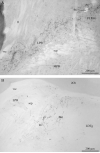
Caudal mesencephalic nuclei. A: Bregma –5.02. Medium-sized fibers stretching from the VLPAG to the LPB and sparse medium-sized fibers in the MPB. B: Bregma –5.52. Medium-sized fibers in the caudal LPB, LC, and Bar. Note that there are almost no fibers in the adjacent LDTg. Scale bars = 200 μm.
Figure 20
Figure 20
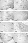
Metencephalon and myelencephalon. A: Bregma –4.36. Small fibers in the ATg at the intersection of the decussation of the superior cerebellar peduncle (xscp) and the tectospinal tract (ts). B: Bregma –4.72. Small fibers at the midline in the MnR at the level of the pons. C: Bregma –5.52. Medium-sized fibers in the IRt; arrows indicate representative fibers. D: Magnification of medium-sized fibers in the Sol from boxed area in E. E: Bregma –6.36. Medium-sized fibers are found in the Sol and in the RMg and GiA. F: Lateral –0.04. Sagittal section near the midline. Dense medium-sized fibers are found in the Sol. Scattered medium-sized fibers are seen in the medullary raphe nuclei, including ROb, RMg, and RPa. G: Magnification of medium-sized fibers in Sol from top boxed area in F. H: Magnification of medium-sized fibers in the raphe nuclei from bottom boxed area in F. I: Lateral 1.2. Medium-sized fibers are seen in the LRt. J: Lateral 1.56. Medium-sized fibers are seen in the Sp5C. Scale bars = 200 μm in A,D,G,H,J; 100 μm in B; 500 lm in C,E,F,I.
Figure 20
Figure 20
Metencephalon and myelencephalon. A: Bregma –4.36. Small fibers in the ATg at the intersection of the decussation of the superior cerebellar peduncle (xscp) and the tectospinal tract (ts). B: Bregma –4.72. Small fibers at the midline in the MnR at the level of the pons. C: Bregma –5.52. Medium-sized fibers in the IRt; arrows indicate representative fibers. D: Magnification of medium-sized fibers in the Sol from boxed area in E. E: Bregma –6.36. Medium-sized fibers are found in the Sol and in the RMg and GiA. F: Lateral –0.04. Sagittal section near the midline. Dense medium-sized fibers are found in the Sol. Scattered medium-sized fibers are seen in the medullary raphe nuclei, including ROb, RMg, and RPa. G: Magnification of medium-sized fibers in Sol from top boxed area in F. H: Magnification of medium-sized fibers in the raphe nuclei from bottom boxed area in F. I: Lateral 1.2. Medium-sized fibers are seen in the LRt. J: Lateral 1.56. Medium-sized fibers are seen in the Sp5C. Scale bars = 200 μm in A,D,G,H,J; 100 μm in B; 500 lm in C,E,F,I.
Figure 21
Figure 21
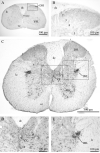
Spinal cord. A: Cervical 6 (approxiamate segmental location). Sparse to scattered fibers are observed in layer 10 around the CC and layers 1, 2, and 3 of the dorsal horn through most of the cervical spinal cord. B: Magnification of medium-sized fibers in dorsal horn from boxed area in A. C: Thoracic 1. In the upper thoracic cord, medium-sized fibers increase in layer 10, and some project laterally to the IML. D: Magnification of medium-sized fibers around CC from midline box in C. E: Magnification of medium-sized fibers in the IML from right box in C. F: Thoracic 11. Medium-sized fibers are again found only around the CC and in the dorsal horn in the caudal thoracic cord. G: Lumbar 5. more fibers are found in dorsal horn in the lumbar cord than in other cord regions. H: Sacral 1. Moderate medium-sized fibers are found in layer 10 from L6 to S2. Some fibers project laterally into the SPSy. I: Magnification of medium-sized fibers in layer 10 and SDCom of the sacral cord from box at midline in H. J: Magnification of medium-sized fibers in the SPSy in the sacral cord from left box in H. Scale bars = 500 μm in A,C,F–H; 100 μm in B,D,E,I,J.
Figure 21
Figure 21
Spinal cord. A: Cervical 6 (approxiamate segmental location). Sparse to scattered fibers are observed in layer 10 around the CC and layers 1, 2, and 3 of the dorsal horn through most of the cervical spinal cord. B: Magnification of medium-sized fibers in dorsal horn from boxed area in A. C: Thoracic 1. In the upper thoracic cord, medium-sized fibers increase in layer 10, and some project laterally to the IML. D: Magnification of medium-sized fibers around CC from midline box in C. E: Magnification of medium-sized fibers in the IML from right box in C. F: Thoracic 11. Medium-sized fibers are again found only around the CC and in the dorsal horn in the caudal thoracic cord. G: Lumbar 5. more fibers are found in dorsal horn in the lumbar cord than in other cord regions. H: Sacral 1. Moderate medium-sized fibers are found in layer 10 from L6 to S2. Some fibers project laterally into the SPSy. I: Magnification of medium-sized fibers in layer 10 and SDCom of the sacral cord from box at midline in H. J: Magnification of medium-sized fibers in the SPSy in the sacral cord from left box in H. Scale bars = 500 μm in A,C,F–H; 100 μm in B,D,E,I,J.

References

    1. Abrahamson EE, Moore RY. Suprachiasmatic nucleus in the mouse: retinal innervation, intrinsic organization and efferent projections. Brain Res. 2001;916:172–191. - PubMed
    1. Aguilera G, Subburaju S, Young S, Chen J. The parvo-cellular vasopressinergic system and responsiveness of the hypothalamic pituitary adrenal axis during chronic stress. Prog Brain Res. 2008;170:29–39. - PMC - PubMed
    1. Albers HE, Rowland CM, Ferris CF. Arginine-vasopressin immunoreactivity is not altered by photoperiod or gonadal hormones in the Syrian hamster (Mesocricetus auratus). Brain Res. 1991;539:137–142. - PubMed
    1. Alelu-Paz R, Gimenez-Amaya JM. The mediodorsal thalamic nucleus and schizophrenia. J Psychiatry Neurosci. 2008;33:489–498. - PMC - PubMed
    1. Andresen MC, Kunze DL. Nucleus tractus solitarius—gateway to neural circulatory control. Annu Rev Physiol. 1994;56:93–116. - PubMed

Publication types

Substances