

Protein nanocapsules containing doxorubicin as a pH-responsive delivery system
- PMID: 21456086
- PMCID: PMC3118673
- DOI: 10.1002/smll.201002242
Protein nanocapsules containing doxorubicin as a pH-responsive delivery system
Abstract
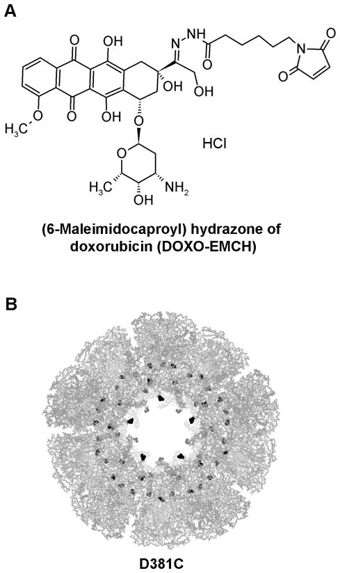
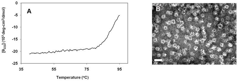
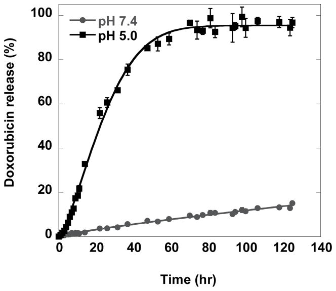
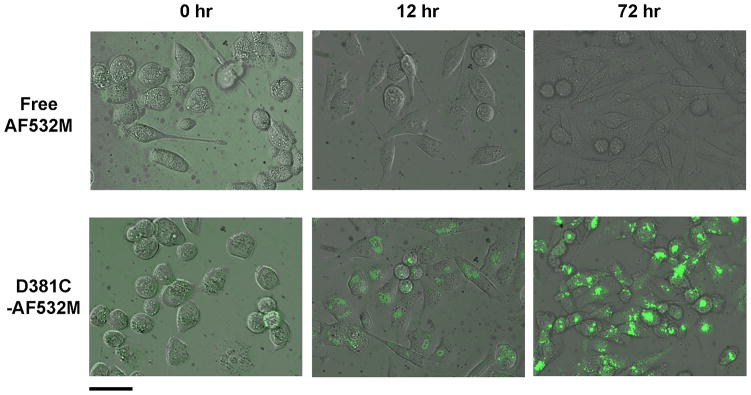
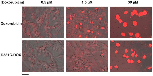
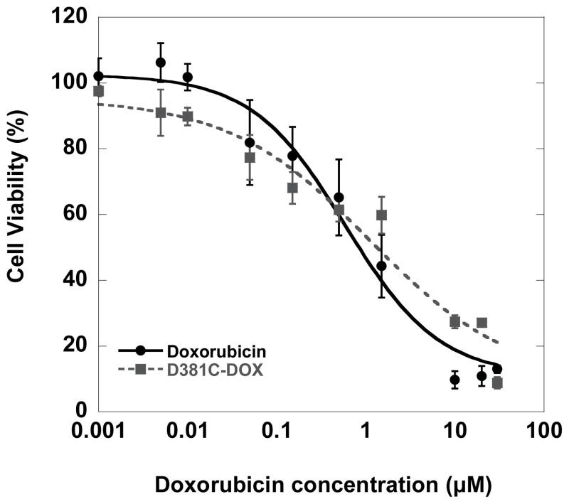
The E2 component of pyruvate dehydrogenase is engineered to form a caged, hollow dodecahedral protein assembly, and the feasibility of this scaffold to be used as a drug delivery system is examined by introducing cysteines to the internal cavity (D381C). The fluorescent dye Alexa Fluor 532 (AF532M) and the antitumor drug doxorubicin are coupled to this internal cavity through maleimides on the guest molecules. The viruslike particle's structure and stability remain intact after binding of the molecules within the interior of the nanocapsule. The pH-dependent hydrolysis of a hydrazone linkage to doxorubicin allows 90% drug release from the D381C scaffold within 72 h at pH 5.0. Fluorescence microscopy of MDA-MB-231 breast cancer cells indicates significant uptake of the D381C scaffold incorporating AF532M and doxorubicin, and suggests internalization of the nanoparticles through endocytosis. It is observed that the protein scaffold does not induce cell death, but doxorubicin encapsulated in D381C is indeed cytotoxic, yielding an IC(50) of 1.3 ± 0.3 μM. While the majority of particulate-based drug delivery strategies encapsulates drugs within polymeric nanoparticles, these results show the potential for using macromolecular protein assemblies. This approach yields a promising new opportunity for designing highly defined nanomaterials for therapeutic delivery.
Copyright © 2011 WILEY-VCH Verlag GmbH & Co. KGaA, Weinheim.
Figures

References
-
- Nie SM, Xing Y, Kim GJ, Simons JW. Annual Review of Biomedical Engineering. 2007;9:257. - PubMed
-
- Ferrari M. Nature Reviews Cancer. 2005;5:161. - PubMed
-
- Doshi N, Mitragotri S. Advanced Functional Materials. 2009;19:3843.
-
- Jiang W, Kim BYS, Rutka JT, Chan WCW. Nature Nanotechnology. 2008;3:145. - PubMed
Publication types
MeSH terms
Substances
Grants and funding
LinkOut - more resources
Full Text Sources
Other Literature Sources
Miscellaneous
