

Review
doi: 10.1021/cr100002u.
Epub 2011 Apr 1.
Genetically encodable fluorescent biosensors for tracking signaling dynamics in living cells
Affiliations
- PMID: 21456512
- PMCID: PMC3092831
- DOI: 10.1021/cr100002u
Item in Clipboard
Review
Genetically encodable fluorescent biosensors for tracking signaling dynamics in living cells
Chem Rev.
.
No abstract available
Figures

Structure of A. victoria GFP. A. victoria GFP showing the dimensions of the protein, the intrinsically-derived p-HBI chromophore and several key residues surrounding the chromophore (image generated using PyMOL open access and PDB ID: 1w7s).

Chromophore formation and ESPT. (A) The most widely accepted mechanism of A. victoria GFP chromophore formation. (B) Proposed mechanism of ESPT for A. victoria GFP (based on ). Both the weakly-fluorescent neutral species (A state) and the highly-fluorescent phenolate anion (B state) are shown, along with their respective excitation and emission maxima (Excitation; Emission).

Chromophore structures of representative FP color variants within each spectral class. The conjugated ring structure of each chromophore is colored according to its emission profile. Excitation and emission maxima are indicated as in Figure 2b.

Genetically-targetable chemical labeling methods. Cartoon depicting several genetically-encodable fluorescent labeling methods that can be applied to biosensor development. The size of each tag is drawn roughly to scale relative to one another and to a FP family member (yellow cylinder). (A) The tetracysteine/biarsenical system. (B) The FAP/fluorogen system. (C) The SNAP-tag/BG system. Note that in the case of (A) and (B), the ligand is virtually non-fluorescent prior to association with the tag.

Genetically-encodable fluorescent tools for studying protein expression/dynamics. (A) When placed under the control of a promoter-of-interest (POI), the maturation of DsRed-Timer from green to red, which occurs over a known period of time, provides information about the timing of promoter activation and attenuation. (B) Fluorescent highlighters composed of a protein-of-interest (blue ovals) and a PI-FP family member (cylinders) allow the movement of sub-populations of tagged proteins to be tracked over time. Here, irradiation with UV-violet light (lightning bolt) within a defined cellular region converts PA-GFP from a non-fluorescent state to a highly fluorescent form, allowing the movement of activated protein-PA-GFP chimeras to be tracked over time.

Fluorescent biosensors to probe biochemical changes within the cellular environment. (A) Localization-based probes for studying PtdIns/lipid dynamics. This example depicts the re-localization of a PtdIns(3,4,5)P3 probe based on the PH domain of Akt (pink pacman) tagged with GFP (green cylinder) from the cytosol to the plasma membrane following generation of PtdIns(3,4,5)P3 by PI3K. (B) A pH/halide sensor based on EYFP. Increases in intracellular pH and/or halide ion concentration (red ball) stabilize the neutral chromophore species, leading to a reduction in fluorescence. (C) Basic design of a single FP-based biosensor. In the example shown, in the “off” state, the sensor domain induces strain on the architecture of an attached FP reporter unit that reduces the fluorescence intensity of the FP. Upon ligand binding (blue circles), the sensor domain undergoes a conformational change that relieves the strain, leading to increased fluorescence in the “on” state (e.g. Camgaroo-2 and HyPer). Alternatively, conformational changes in the sensor domain can alter the excitation and/or emission profile of the FP reporter unit, permitting ratiometric measurements (e.g. ratiometric pericam). (D) Basic design of a FRET-based biosensor. Conformational changes in the sensor domain caused by ligand binding or post-translational modification alter the distance and/or orientation of the attached FP FRET pair, resulting in changes in the efficiency of energy transfer between them (blue arrow).

Origins of a molecular switch. A molecular switch can be generated by either (A) a conformational change intrinsic to a protein or protein domain or (B) by an engineered conformational change driven by interactions between a receiver module (gray block) and a switching module (green block). In each case, the conformation change is converted to a change in FRET efficiency (blue arrow) by altering the distance and/or orientation of the attached FP reporter units (cylinders). Alternatively, a single FP may serve as the reporter unit, as in Figure 6c.
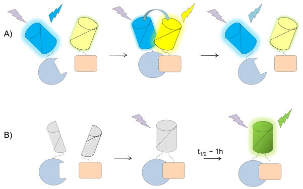
Fluorescent reporters to track protein-protein interactions. (A) FRET-based interaction sensors, which use the association of fluorescently-tagged proteins to change the distance and/or orientation of a FRET pair, allow reversible interactions to be monitored in real-time. (B) Meanwhile, BiFC-based interaction sensors, which are essentially irreversible, use the association of proteins tagged with complementary halves of split FP molecules to promote fluorophore formation. Though the relatively slow maturation of the fluorophore prevents BiFC-based probes from measuring protein-protein interactions in real-time, the large dynamic range of these sensors allows interactions to be detected at protein concentrations that do not dramatically alter the cellular context in which the interactions occur.
References
Publication types
MeSH terms
Substances
Grants and funding
LinkOut - more resources
Full Text Sources
Other Literature Sources
