Review
. 2011:34:259-88.
doi: 10.1146/annurev-neuro-061010-113720.

The cognitive neuroscience of human memory since H.M

Affiliations
Review

The cognitive neuroscience of human memory since H.M

Larry R Squire et al. Annu Rev Neurosci. 2011.

Abstract

Work with patient H.M., beginning in the 1950s, established key principles about the organization of memory that inspired decades of experimental work. Since H.M., the study of human memory and its disorders has continued to yield new insights and to improve understanding of the structure and organization of memory. Here we review this work with emphasis on the neuroanatomy of medial temporal lobe and diencephalic structures important for memory, multiple memory systems, visual perception, immediate memory, memory consolidation, the locus of long-term memory storage, the concepts of recollection and familiarity, and the question of how different medial temporal lobe structures may contribute differently to memory functions.


Figures

Figure 1
Figure 1
Left column Magnetic resonance images arranged from rostral (a) to caudal (c) through the temporal lobe of patient H.M. (in 1993 at age 67) and a 66-year-old healthy male (right). The comparison brain illustrates the structures that appear to have been removed during H.M.’s surgery in 1953. The lesion was bilaterally symmetrical, extending caudally 5.4 cm on the left side and 5.1 cm on the right. The full caudal extent of abnormal tissue is not illustrated. The damage included medial temporal polar cortex, most of the amygdaloid complex, virtually all the entorhinal cortex, and approximately the rostral half of the hippocampal region (dentate gyrus, hippocampus, and subicular complex). The perirhinal cortex was substantially damaged except for its ventrocaudal aspect. The more posterior parahippocampal cortex (areas TF and TH, not shown here) was largely intact. Adapted from Corkin et al. (1997) with permission from the Society for Neuroscience.
Figure 2
Figure 2
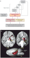
(a) Schematic view of the medial temporal lobe memory system for declarative memory, which is composed of the hippocampus and the perirhinal, entorhinal, and parahippocampal cortices. In addition to the connections shown here, there are also weak projections from the perirhinal and parahippocampal cortices to the CA1-subiculum border. (b) Ventral view of a human brain (upper left), monkey brain (upper right), and a lateral view of a rat brain (lower center). The major cortical components of the medial temporal lobe are highlighted and outlined. The hippocampus is not visible from the surface and in the human lies beneath the cortex of the medial temporal lobe. Its anterior extent lies below the posterior entorhinal (red ) and perirhinal ( purple) cortices, and the main body of the hippocampus lies beneath the parahippocampal cortex. In the rat, the parahippocampal cortex is termed postrhinal cortex. Abbreviations: EC, entorhinal cortex; PH, parahippocampal cortex (dark yellow); Por, postrhinal cortex; PR, perirhinal cortex.
Figure 3
Figure 3
Intact working memory and impaired long-term memory. (a) The number of trials needed to succeed at each string length for patient H.M. and controls. H.M. could not succeed at repeating back 7 digits even after 25 attempts with the same string. (b) The number of trials needed to learn the locations of different numbers of objects for patient G.P. and controls. G.P. could not reproduce the locations of four objects, even after 10 attempts with the same display (panel a adapted from Drachman & Arbit 1966, with permission from the American Medical Association, and panel b adapted from Jeneson et al. 2010).
Figure 4
Figure 4
(a) Participants copied the Rey-Osterrieth figure illustrated in the small box in the upper left and 10–15 min later, without forewarning, tried to reproduce it from memory. The reproduction by a representative control is shown below the target figure. The left panel also shows the reproduction by patient R.B., who had histologically identified lesions of the CA1 field of the hippocampus (Zola-Morgan et al. 1986). Patient E.P., who had large medial temporal lobe lesions, did not recall copying a figure and declined to guess. The right section shows reproductions by seven patients with circumscribed damage to the hippocampus. Panels b and c show scores for the same seven patients (H) and 13 controls on the autobiographical memory interview, childhood portion (Kopelman et al. 1989). These findings suggest that patients who fail to produce any of the complex figure (like E.P.) or who are deficient at producing either remote semantic memories (A, maximum score, 21) or remote autobiographical events (B, maximum score, 9) will prove to have damage beyond the hippocampus. Indeed, even E.P. with his large lesions limited mainly to the medial temporal lobes, obtained maximal scores on these two tests (21/21 and 9/9).
Figure 5
Figure 5
Individual recognition (a) and recall scores (b) for hippocampal patients (n = 7) and healthy controls (n = 8) from Manns et al. (2003a). When the patient scores for recognition and recall are converted to z-scores based on the mean and standard deviation of the corresponding control scores, the recognition deficit (−1.59) is statistically indistinguishable from the recall deficit (−1.81), p > 0.60. d′ = discriminability.
Figure 6
Figure 6
Symmetrical (a) and asymmetrical (b) receiver operating characteristic (ROC) plots with hypothetical data shown as filled red circles. The axis of symmetry is the negative diagonal (dashed gray line), and chance performance is indicated by the positive diagonal (solid blue line). The symmetrical ROC (a) reflects relatively weak memory (the data fall close to the positive diagonal), and the asymmetrical ROC (b) reflects stronger memory (the data fall farther from the positive diagonal).

References

    1. Aggleton JP, Vann SD, Denby C, Dix S, Mayes AR, et al. Sparing of the familiarity component of recognition memory in a patient with hippocampal pathology. Neuropsychologia. 2005;43:810–23. - PubMed
    1. Atkinson RC, Juola JF. Search and decision processes in recognition memory. In: Krantz DH, Atkinson RC, Suppes P, editors. Contemporary Developments in Mathematical Psychology. San Francisco: Freeman; 1974. pp. 243–90.
    1. Baddeley A. Working memory: looking back and looking forward. Nat Rev Neurosci. 2003;4:829–39. - PubMed
    1. Barense MD, Gaffan D, Graham KS. The human medial temporal lobe processes online representations of complex objects. Neuropsychologia. 2007;45:2963–74. - PubMed
    1. Barton JJS, Cherkasova M. Face imagery and its relation to perception and covert recognition in prosopagnosia. Neurology. 2003;61:220–25. - PubMed

Publication types