

Review
doi: 10.1186/ar3251.
Abnormalities of T cell signaling in systemic lupus erythematosus
Affiliations
- PMID: 21457530
- PMCID: PMC3132009
- DOI: 10.1186/ar3251
Item in Clipboard
Review
Abnormalities of T cell signaling in systemic lupus erythematosus
Arthritis Res Ther.
.
Abstract
Systemic lupus erythematosus (SLE) is an autoimmune disease resulting from a loss of tolerance to multiple self antigens, and characterized by autoantibody production and inflammatory cell infiltration in target organs, such as the kidneys and brain. T cells are critical players in SLE pathophysiology as they regulate B cell responses and also infiltrate target tissues, leading to tissue damage. Abnormal signaling events link to defective gene transcription and altered cytokine production, contributing to the aberrant phenotype of T cells in SLE. Study of signaling and gene transcription abnormalities in SLE T cells has led to the identification of novel targets for therapy.
Figures
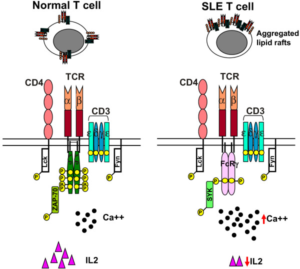
Schematic showing the T cell receptor signaling architecture in normal and systemic lupus erythematosus T cells. SLE, systemic lupus erythematosus; TCR, T cell receptor.

Schematic showing transcription factors involved in IL2 production in T cells. AP1, activated protein 1; CAMKIV, calcium/calmodulin-dependent kinase IV; CREB, cAMP response element-binding; CREM, cAMP response element modulator; MAPK, mitogen-activated protein kinase; NFAT, nuclear factor of activated T cells; PKC, protein kinase C; PP, protein phosphatase.

Schematic showing the CD3ζ gene. Genomic DNA with eight exons (top), the mRNA with a full-length 906-bp 3' UTR (WT; middle) and the 344-bp alternatively spliced (AS) 3' UTR variant (bottom). SLE T cells express increased amounts of the unstable AS splice variant relative to the stable WT isoform.
References
-
- Krishnan S, Nambiar MP, Warke VG, Fisher CU, Mitchell J, Delaney N, Tsokos GC. Alterations in lipid raft composition and dynamics contribute to abnormal T cell responses in systemic lupus erythematosus. J Immunol. 2004;172:7821–7831. - PubMed
Publication types
MeSH terms
Grants and funding
LinkOut - more resources
Full Text Sources
Other Literature Sources
Medical
