. 2011 Apr 1;4(1):8.
doi: 10.1186/1755-8166-4-8.

A rare case of t(11;22) in a mantle cell lymphoma like B-cell neoplasia resulting in a fusion of IGL and CCND1: case report

Affiliations

A rare case of t(11;22) in a mantle cell lymphoma like B-cell neoplasia resulting in a fusion of IGL and CCND1: case report

Cristiano Krings Rocha et al. Mol Cytogenet. .

Abstract

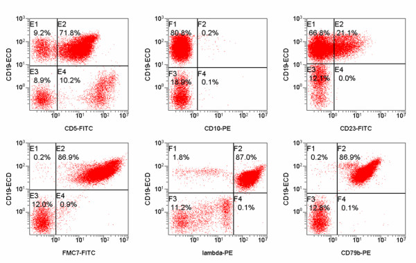
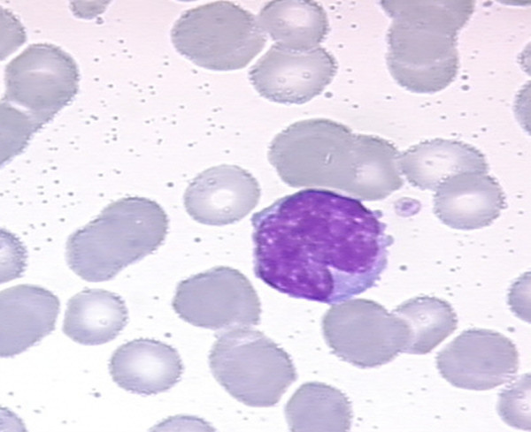
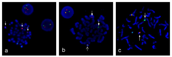
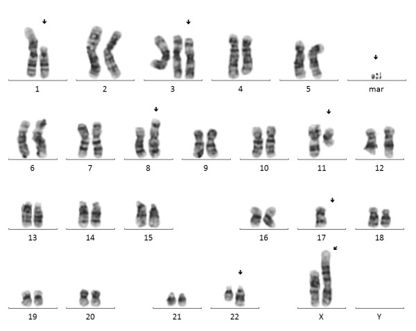
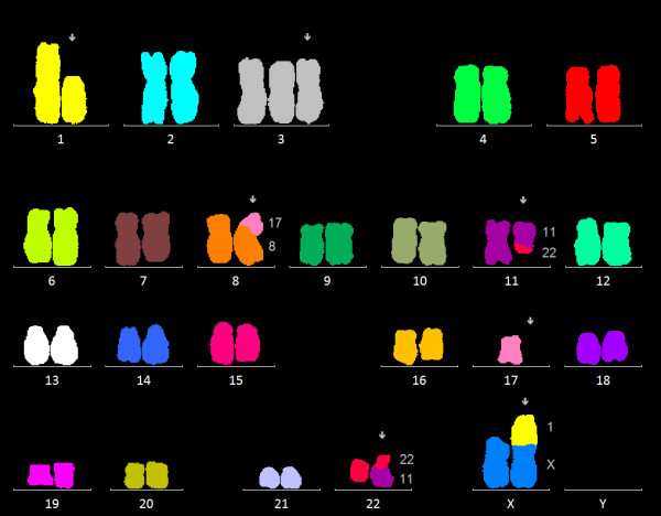
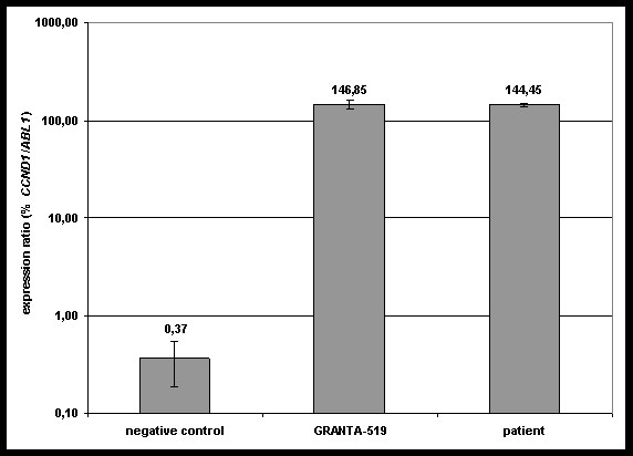
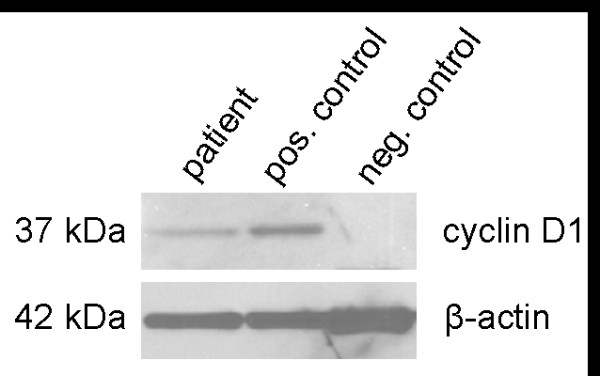
The chromosomal translocation (11;14)(q13;q32) rearranging the locus for cyclin D1 (CCND1) to that of the immunoglobulin heavy chain (IGH) can be found in virtually all cases of mantle cell lymphoma (MCL), while other CCND1 translocations are extremely rare. As CCND1 overexpression and activation is a hallmark of MCL it is regarded as a central biological mechanism in the development and maintenance of this disease.Here we present a patient initially diagnosed with chronic lymphocytic leukemia (CLL) where chromosome banding analysis revealed, among other aberrations, a translocation (11;22)(q13;q11.2). We show by fluorescence in situ hybridization (FISH) analysis that on chromosome 22 the immunoglobulin light chain lambda (IGL) is involved in this cytogenetic aberration. Additionally, we demonstrate the resulting overexpression of CCND1 on the RNA and protein level, thereby consolidating the new diagnosis of a MCL-like B-cell neoplasia. Summing up, we described a rare case of t(11;22)(q13;q11.2) in a MCL-like neoplasia and showed that this aberration leads to an overexpression of CCND1 which is regarded as a key biological feature in MCL. This case underlines the importance of cytogenetic analyses especially in atypical cases of B cell lymphomas.


Figures

Figure 1
Figure 1
Flow cytometry results. Flow cytometric graphs showing positivity for CD5, CD79b, FMC7, surface immunoglobulin lambda and negativity for CD23 and CD10.
Figure 2
Figure 2
Morphology of patient's leukaemic cells. Medium-sized lymphoid cell with lightly basophilic cytoplasm and medium-sized irregular nucleus with dispersed chromatin.
Figure 3
Figure 3
Metaphase and interphase FISH. (a) FISH showing 3 signals for CCND1 due to a translocation at the CCND1 locus on 11(q13). CCND1 on 11(q13) is marked in red, IGH on 14(q32) is marked in green. (b) FISH confirming IGL rearrangement with an IGL dual-color breakapart probe. Broad arrows show separated signals for the IGL proximal region in red and for the distal region in green. The open arrow shows a fusion signal for the normal chromosome 22. (c) FISH with a CCND1/IGL tri-color, dual-fusion probe, showing the IGL/CCND1 translocation. CCND1 on 11(q13) marked in Spectrum Orange and Spectrum Green, IGL on 22(q11.2) marked in Spectrum Aqua. The broad arrow shows the derivative chromosome 22 and the open arrow the derivative chromosome 11.
Figure 4
Figure 4
Conventional chromosome analysis. Complex aberrant karyogram of patient showing the karyotype 46, X, der(X)t(X;1)(p22.1;p21), del(1)(p21), +3, der(8)t(8;17)(p21;q21), t(11;22)(q13;q11.2), -17, 3dmin.
Figure 5
Figure 5
mFISH analysis. Multicolor FISH analysis confirmed the deletion on chromosome 1, the trisomy 3, the unbalanced translocation (8;17), the balanced translocation (11;22), the loss of one chromosome 17 and the unbalanced translocation (X;1).
Figure 6
Figure 6
Quantitative real-time RT-PCR results. The overexpression of CCND1 could be clearly detected for the patient in comparison to GRANTA-519 CCND1-positive cells. Expression ratios are given as %CCND1/ABL1 in log-scale on the y axis.
Figure 7
Figure 7
Western blot analysis of CCND1 expression. The overexpression of cyclin D1 in our patient is clearly visible (left panel) as well as in GRANTA-519 cells with t(11;14)(q13;q32) (middle panel) compared to a healthy donor (right panel).

References

    1. Morton LM, Wang SS, Devesa SS, Hartge P, Weisenburger DD, Linet MS. Lymphoma incidence patterns by WHO subtype in the United States, 1992-2001. Blood. 2006;107:265–276. doi: 10.1182/blood-2005-06-2508. - DOI - PMC - PubMed
    1. Jares P, Campo E. Advances in the understanding of mantle cell lymphoma. Br J Haematol. 2008;142:149–165. doi: 10.1111/j.1365-2141.2008.07124.x. - DOI - PubMed
    1. Pileri SA, Falini B. Mantle cell lymphoma. Haematologica. 2009;94:1488–1492. doi: 10.3324/haematol.2009.013359. - DOI - PMC - PubMed
    1. Komatsu H, Yoshida K, Seto M, Iida S, Aikawa T, Ueda R. et al. Overexpression of PRAD1 in a mantle zone lymphoma patient with a t(11;22)(q13;q11) translocation. Br J Haematol. 1993;85:427–429. doi: 10.1111/j.1365-2141.1993.tb03194.x. - DOI - PubMed
    1. Quintanilla-Martinez L, Slotta-Huspenina J, Koch I, Klier M, Hsi ED, de LL. et al. Differential diagnosis of cyclin D2+ mantle cell lymphoma based on fluorescence in situ hybridization and quantitative real-time-PCR. Haematologica. 2009;94:1595–1598. doi: 10.3324/haematol.2009.010173. - DOI - PMC - PubMed