. 2011 Jul 1;51(1):171-8.
doi: 10.1016/j.freeradbiomed.2011.03.025. Epub 2011 Mar 30.

NADPH-oxidase activation and cognition in Alzheimer disease progression

Affiliations

NADPH-oxidase activation and cognition in Alzheimer disease progression

Mubeen A Ansari et al. Free Radic Biol Med. .

Abstract

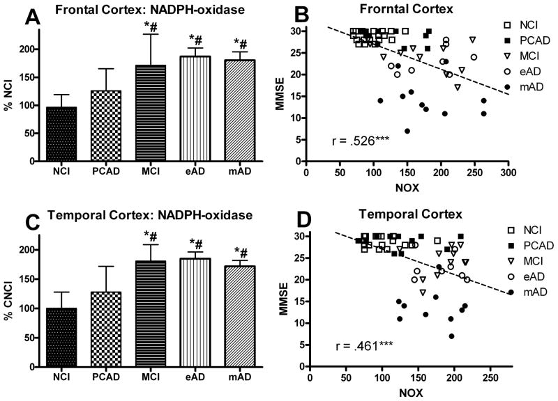
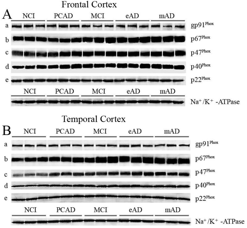
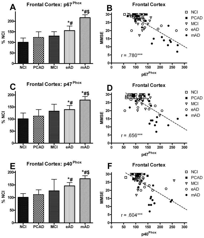
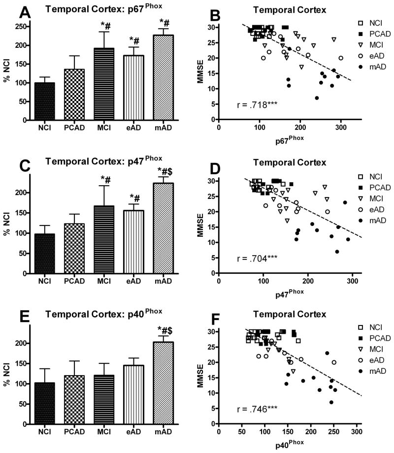
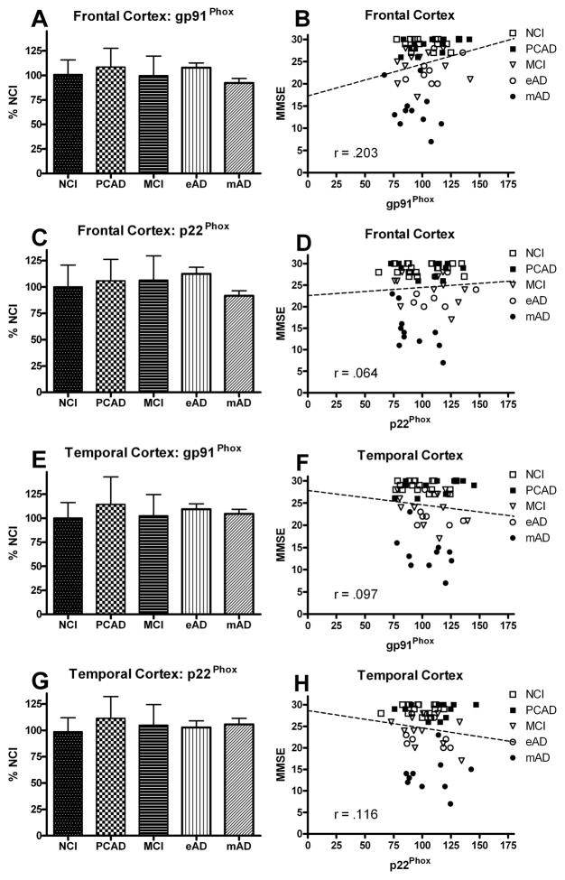
Superoxide production via NADPH-oxidase (NOX) has been shown to play a role in a variety of neurological disorders, including Alzheimer disease (AD). To improve our understanding of the NOX system and cognitive impairment, we studied the various protein components of the phagocytic isoform (gp91(phox), or NOX2) in the frontal and temporal cortex of age- and postmortem-matched samples. Individuals underwent antemortem cognitive testing and postmortem histopathologic assessment to determine disease progression and assignment to one of the following groups: no cognitive impairment (NCI), preclinical AD, mild cognitive impairment (MCI), early AD, and mild-to-moderate AD. Biochemical methods were used to determine overall NOX activity as well as levels of the various subunits (gp91(phox), p67(phox), p47(phox), p40(phox), and p22(phox)). Overall enzyme activity was significantly elevated in the MCI cohort in both cortical regions compared to the NCI cohort. This activity level remained elevated in the AD groups. Only the NOX cytosolic subunit proteins (p67(phox), p47(phox), and p40(phox) ) were significantly elevated with disease progression; the membrane-bound subunits (gp91(phox) and p22(phox)) remained stable. In addition, there was a robust correlation between NOX activity and the individual's cognitive status such that as the enzyme activity increased, cognitive performance decreased. Collectively, these data show that upregulated NADPH-oxidase in frontal and temporal cortex suggests that increases in NOX-associated redox pathways might participate in early pathogenesis and contribute to AD progression.


Figures

FIG. 1
FIG. 1
Levels of NADPH-oxidase (NOX) activity analyzed in human (A) frontal and (C) temporal cortex from individuals classified as NCI, PCAD, MCI, eAD, and mAD. Bars represent group mean ± SD. *p < 0.01 versus NCI, #p < 0.01 versus PCAD. Scatter plots show the association between NOX activity and the subject’s score on the Mini Mental Status Exam (B) FC and (D) TC. Line used to represent the direction of the association and does not indicate a line of regression. *** p < 0.001 Spearman’s rho. In both FC and TC, individuals with increased NADPH-oxidase activity had lower MMSE scores.
FIG. 2
FIG. 2
Representative Western-blots demonstrating changes in NADPH-oxidase protein subunits in the isolated membrane fractions from NCI, PCAD, MCI, eAD, and mAD subjects in frontal cortex (A), and temporal cortex (B) as a function of disease progression. The values were normalized with standard membrane marker Na+/K+ -ATPase. Isolated membrane fractions from all subjects were processed for immunobloting followed by Western-blot.
FIG. 3
FIG. 3
Levels of cytosolic protein subunits of NADPH-oxidase (p67Phox, p47Phox, p40Phox) (A,C,E) analyzed in membrane fractions of the frontal cortex from individuals classified as NCI, PCAD, MCI, eAD, and mAD. Values are plotted as percent change from the levels observed in the NCI cohort. Bars represent group mean ± SD. *p < 0.01 versus NCI, #p < 0.01 versus PCAD, $ p < 0.01 versus MCI. Scatter plots show the relationship between the individual’s score on the MMSE and the levels of the different cytosolic subunits (B,D,E). Each point represents an individual subject. Line used to represent the direction of the association and does not indicate a line of regression. *** p < 0.001 Spearman’s rho
FIG. 4
FIG. 4
Levels of cytosolic protein subunits of NADPH-oxidase (p67Phox, p47Phox, p40Phox) (A,C,E) analyzed in membrane fractions of the temporal cortex from individuals classified as NCI, PCAD, MCI, eAD, and mAD. Values are plotted as percent change from the levels observed in the NCI cohort. Bars represent group mean ± SD. *p < 0.01 versus NCI, #p < 0.01 versus PCAD, $ p < 0.01 versus MCI. Scatter plots show the relationship between the individual’s score on the MMSE and the levels of the different cytosolic subunits (B,D,E). Each point represents an individual subject. Line used to represent the direction of the association and does not indicate a line of regression. *** p < 0.001 Spearman’s rho
FIG. 5
FIG. 5
Levels of membrane subunit proteins of NADPH-oxidase (gp91Phox, and p22Phox) in the frontal (A,C) and temporal (E,G) cortex from individuals classified as NCI, PCAD, MCI, eAD, and mAD. Values are plotted as percent change from the levels observed in the NCI cohort. Bars represent group mean ± SD. Scatter plots show the relationship between the individual’s score on the MMSE and the levels of the different membrane subunits (B,D,F,H). Each point represents an individual subject. Line used to represent the direction of the association and does not indicate a line of regression.

References

    1. Nelson PT, Braak H, Markesbery WR. Neuropathology and cognitive impairment in Alzheimer disease: a complex but coherent relationship. J Neuropathol Exp Neurol. 2009;68:1–14. - PMC - PubMed
    1. Scheff SW, Price DA. Alzheimer’s disease-related alterations in synaptic density: neocortex and hippocampus. J Alzheimers Dis. 2006;9:101–115. - PubMed
    1. Petersen RC, Morris JC. Mild cognitive impairment as a clinical entity and treatment target. Arch Neurol. 2005;62:1160–1163. discussion 1167. - PubMed
    1. Winblad B, Palmer K, Kivipelto M, Jelic V, Fratiglioni L, Wahlund LO, Nordberg A, Backman L, Albert M, Almkvist O, Arai H, Basun H, Blennow K, de Leon M, DeCarli C, Erkinjuntti T, Giacobini E, Graff C, Hardy J, Jack C, Jorm A, Ritchie K, van Duijn C, Visser P, Petersen RC. Mild cognitive impairment--beyond controversies, towards a consensus: report of the International Working Group on Mild Cognitive Impairment. J Intern Med. 2004;256:240–246. - PubMed
    1. Morris JC, Storandt M, McKeel SW, Rubin eH, Price JL, Grant EA, Berg L. Cerebral amyloid deposition and diffuse plaques in “normal” aging: Evidence for presymptomatic and very mild Alzheimer’s disease. Neurology. 1996;46:707–719. - PubMed

Publication types