. 2011 Apr 22;34(4):479-91.
doi: 10.1016/j.immuni.2011.02.017. Epub 2011 Mar 31.

Structure of a domain-swapped FOXP3 dimer on DNA and its function in regulatory T cells

Affiliations

Structure of a domain-swapped FOXP3 dimer on DNA and its function in regulatory T cells

Hozefa S Bandukwala et al. Immunity. .

Erratum in

  • Immunity. 2011 Apr 22;34(4):627

Abstract

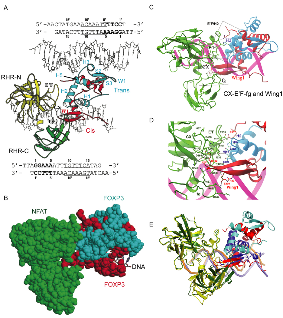
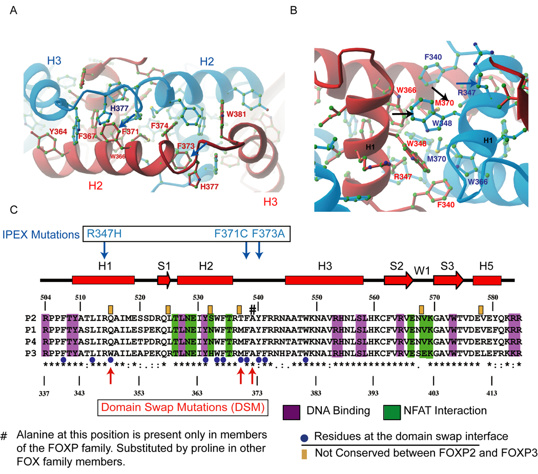
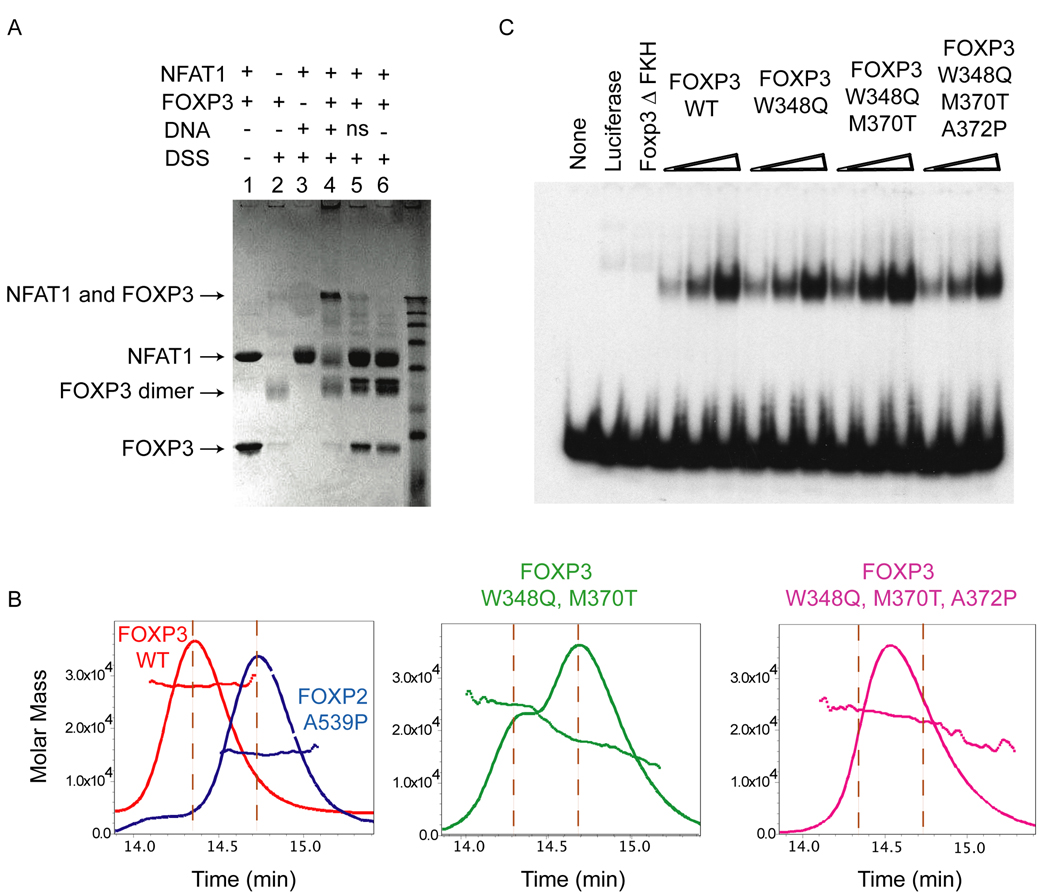
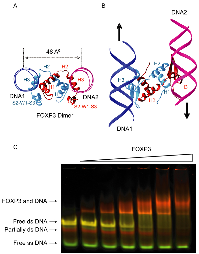
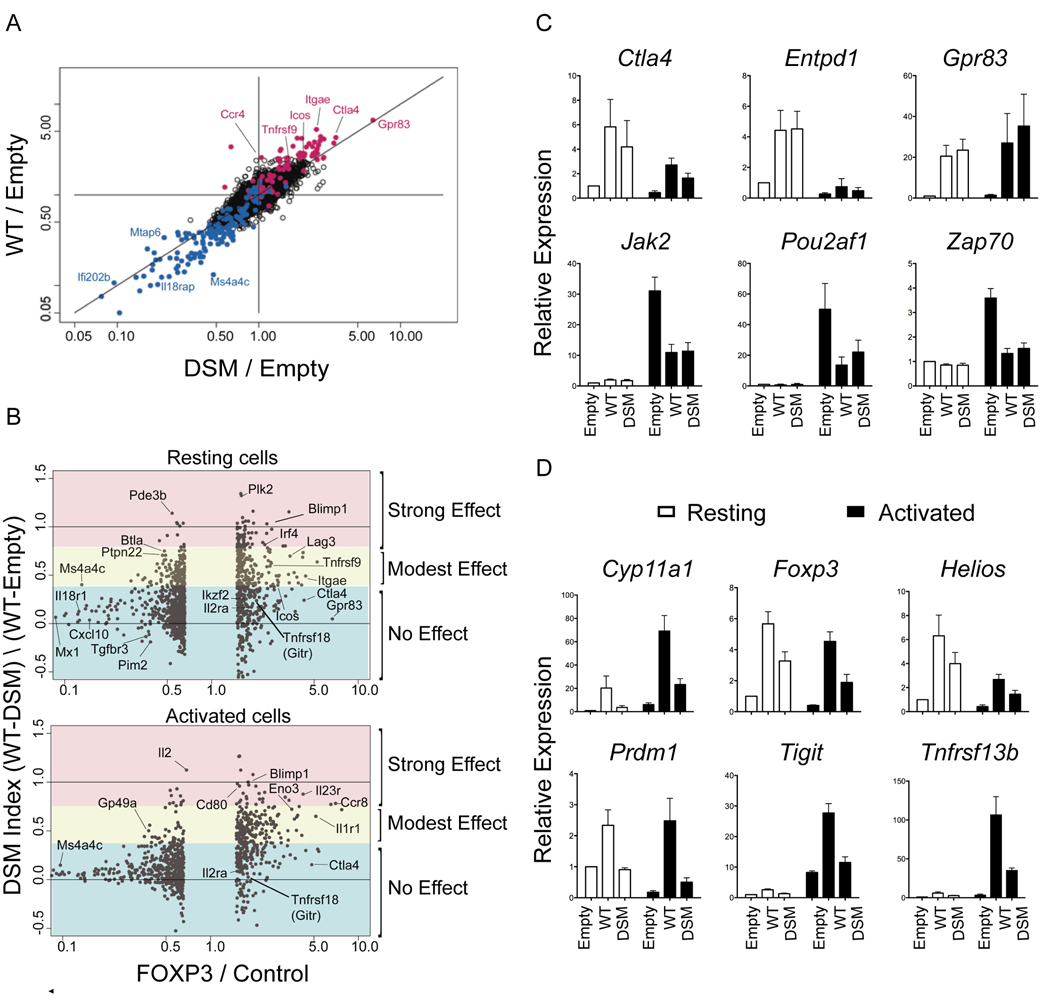
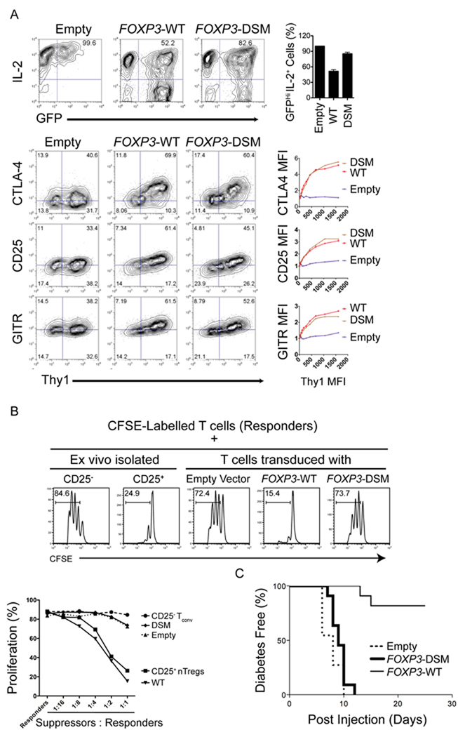
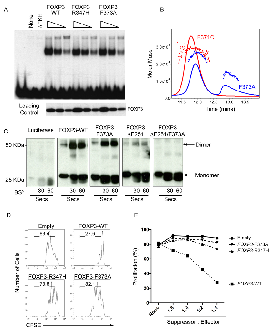
The transcription factor FOXP3 is essential for the suppressive function of regulatory T cells that are required for maintaining self-tolerance. We have solved the crystal structure of the FOXP3 forkhead domain as a ternary complex with the DNA-binding domain of the transcription factor NFAT1 and a DNA oligonucleotide from the interleukin-2 promoter. A striking feature of this structure is that FOXP3 forms a domain-swapped dimer that bridges two molecules of DNA. Structure-guided or autoimmune disease (IPEX)-associated mutations in the domain-swap interface diminished dimer formation by the FOXP3 forkhead domain without compromising FOXP3 DNA binding. These mutations also eliminated T cell-suppressive activity conferred by FOXP3, both in vitro and in a murine model of autoimmune diabetes in vivo. We conclude that FOXP3-mediated suppressor function requires dimerization through the forkhead domain and that mutations in the dimer interface can lead to the systemic autoimmunity observed in IPEX patients.


Figures

Figure 1
Figure 1. Overall structure of the NFAT1:FOXP3:DNA complex
(A) Arrangement of protein and DNA components in the asymmetric unit. The N-terminal (RHR-N: yellow) and C-terminal domains (RHR-C: green) of NFAT1, and the cis- (red) and trans-monomer (cyan) of FOXP3 are shown. The sequence of each DNA molecule (stick model) is shown along its orientation in the crystal. The FOXP3 binding site of the cis-monomer (bottom DNA) and trans-monomer (top DNA) are underlined. The NFAT site is in bold. (B) Protein-protein interactions between NFAT1 and FOXP3. The NFAT1 (green) and FOXP3 dimer (red and cyan) are shown in space-filling model and DNA in stick model (C) Structural elements of each FOXP3 monomer contribute to protein-protein interactions with NFAT1. Helix H2 of the trans-monomer forms an extensive interaction interface with the E’F loop of NFAT1. The Wing1 of the cis-monomer inserts into a groove formed by the CX loop, the C-terminal stem of the E’F loop, and the fg loop of NFAT). (D) Detailed interactions between NFAT and the FOXP3 dimer. Interacting residues are coloured and labelled according to their respective proteins. (E) Superposition of the complex of NFAT1 (yellow), FOXP3 dimer (red and cyan) and DNA (purple) with the complex of NFAT1 (green), FOXP2 monomer (blue) and DNA (orange). The DNA (orange) in the NFAT1:FOXP2:DNA complex is clearly bent whereas DNA (purple) in the NFAT1:FOXP3:DNA complex is nearly straight. The NFAT-binding elements (helix H2 and wing W1) of FOXP2 and the FOXP3 dimer are aligned similarly toward NFAT but with a distinct shift.
Figure 2
Figure 2. The dimerisation interface in the forkhead domain of FOXP3
(A) An interaction network of aromatic residues at the domain-swapped dimer interface present in both FOXP2 and FOXP3 (see Figure S3, top left). The red and blue monomers of FOXP3 are shown with residues coloured according to the monomer to which they belong. The different conformations of His377 in the two monomers are induced by different DNA binding interactions at Site A and Site B (see Figure S2 for details) (B) An additional hydrophobic core (exclusive to FOXP3, see Figure S3, top right) that enhances the stability of the domain-swapped FOXP3 dimer. (C) Sequence alignment of FOXP family. The secondary structural elements are shown above the sequence. Numbering of FOXP2 is shown above whereas that of FOXP3 is shown below. The homology is indicated by standard convention below of the sequence alignment. Residues involved in DNA binding (magenta), NFAT interaction (green), and domain swapping (blue dots) are highlighted. The six residues that differ between FOXP2 and FOXP3 forkhead domains are indicated by orange boxes above the sequence alignment. The blue downward-pointing arrows indicate the positions of IPEX mutations R347H, F371C and F373A that occur at the domain-swap interface, and the red upward arrows and the pound sign depict the position of the structure-guided mutations that we engineered into the interface (see text )
Figure 3
Figure 3. DSM mutations affect dimerization, but do not impair DNA binding by the forkhead domain of FOXP3
(A) Denaturing SDS gel showing formation of a crosslinked FOXP3 dimer after DSS crosslinking (lanes 1 and 2), and sequence-dependent cross-linking of NFAT1 and FOXP3 on the ARRE2 DNA element (lanes 3–6). The composition of each reaction is indicated above the lane, and the concentrations of each component are described in Methods. ns, nonspecific DNA. (B) SEC-MALS analysis of the forkhead domains of WT-FOXP3 and the indicated mutants. FOXP3 exists as a stable dimer in solution and elutes as a single peak (~26 kDa, left panel); the double mutant; W348Q and M370T FOXP3 elutes as two peaks of 26 kDa and 13 kDa representing the dimeric and monomeric forms (middle panel); and the triple DSM mutant; W348Q, M370T and A372P elutes as a single polydisperse peak (right panel). (C) EMSA analysis depicting binding of WT-FOXP3 and indicated mutants to a DNA probe containing optimized FOXP3 binding sequences. The mutations do not impair DNA binding by FOXP3.
Figure 4
Figure 4. The FOXP3 forkhead domain can bring together two FOXP3 DNA binding elements in solution
The domain-swapped FOXP3 dimer (red and cyan) binds two separate DNA molecules (magenta and blue, only backbones are shown in ribbons). (A) View along the DNA axis. (B) View from the top. (C) In-Gel FRET analysis: Pseudo-colored image showing an overlay of the fluorescence of the Cy3-donor (green) and the fluorescence of the Cy5-acceptor (red) fluorophores. Labels to the left indicate the respective DNA/ protein species. Note, unannealed probe (free ss DNA, green) is not bound by FOXP3 forkhead dimer even at high protein concentrations and remains unretarded during electrophoresis.
Figure 5
Figure 5. Mutations that disrupt domain swapping differentially affect regulation of FOXP3 target genes
(A) Gene expression in transfected cells represented as FoldChange versus FoldChange (FcFc) plots comparing the effect of transduction of WT and DSM Foxp3 relative to mock-transfected cells (resting conditions; all microarray profiles performed in duplicate). (B) Changes in gene expression as a function of the DSM mutation represented as DSM index (calculated from expression values, (WT-DSM) / (WT-Empty)) versus FoldChange in expression obtained upon FOXP3 transduction in resting (top) and activated cells (bottom). (C and D) Expression of indicated FOXP3 target genes in WT and DSM transduced T cells as estimated by real-time PCR analysis. Error bars represent the SEM observed in three independent experiments.
Figure 6
Figure 6. The domain-swap interface is required for the function of FOXP3
(A) Representative flow cytometry plots depicting expression of IL-2 (top panel), CTLA-4, GITR and CD25 (bottom panels) in CD4+ T cells transduced with an empty control vector encoding WT or DSM-FOXP3. The X-axis denotes GFP fluorescence, used as a proxy for FOXP3 expression; GFP expression is controlled by an IRES element within the bicistronic RNA encoding the FOXP3 coding sequence. The data are representative of three independent experiments. Bar graphs on right represent percent of IL-2hi cells within the GFP+ population (top panel) and fold increase in MFI of CTLA4 and CD25 in transduced cells (middle and bottom panels respectively). The data are representative of 3 independent experiments. (B) Cells expressing the FOXP3 domain swap mutant show decreased suppressor function in vitro. Top, Representative histograms depicting the CFSE dilution profile of responder CD4+ T cells cultured with either ex-vivo isolated CD4+ CD25+ T cells or transduced FOXP3-expressing cells (suppressors) at a ratio of 1:2 (suppressors:responders). Bottom, graph demonstrating relative proliferation of responder cells cultured with transduced FOXP3-expressing suppressor cells at different suppressor:respondor ratios. The data are representative of two independent experiments. (C) Cells transduced with DSM-FOXP3 fail to prevent induction of diabetes in NOD recipient mice. Neonatal BDC2.5 TCR transgenic NOD mice were injected with T cells retrovirally transduced with WT or DSM-FOXP3 together with Th1 cell effectors generated from BDC2.5 TCR transgenic mice. The graph represents the kinetics of diabetes induction in recipient mice: cells transduced with WT-FOXP3 prevent diabetes induction by the effector Th1 cells, whereas cells transduced with DSM-FOXP3 are ineffective. The data were obtained from three independent experiments.
Figure 7
Figure 7. IPEX mutations that disrupt domain swapping diminish FOXP3 regulatory activity
(A) Forkhead mutations that lie at the domain swap interface do not disrupt DNA binding. EMSA analysis comparing the ability of WT and indicated FOXP3 forkhead mutants to bind DNA. (B) The F373A mutation at the domain swap interface destabilizes dimer formation by the FOXP3 forkhead domain. SEC-MALS analysis of the forkhead domains of FOXP3 F371C and F373A shows that the F371C mutant remains a dimer whereas the F373A mutants elutes as a mixture of monomer and dimer. (C) Denaturing SDS Gel of FOXP3 (aa 182–431) before and after brief crosslining with BS3, showing that FOXP3 contains two dimerisation interfaces in the leucine zipper and the forkhead domain. The combination of the ΔE251 mutation in the leucine zipper and the F373A mutation in the forkhead domain eliminates dimer formation. (D) IPEX mutations that destabilize domain swapping abrogate the ability of FOXP3 to induce regulatory functions. Shown is a representative histogram depicting the CFSE dilution profile of responder cells cultured with transduced WT-FOXP3 or IPEX mutant-expressing cells (suppressors) at a ratio of 1:2 (suppressors:responders). (E), Graph quantitating relative proliferation of CFSE-labelled responder cells cultured with transduced FOXP3-expressing suppressor cells at different suppressor: responder ratios. These data are representative of at least two independent experiments.

References

    1. Bacchetta R, Passerini L, Gambineri E, Dai M, Allan SE, Perroni L, Dagna-Bricarelli F, Sartirana C, Matthes-Martin S, Lawitschka A, et al. Defective regulatory and effector T cell functions in patients with FOXP3 mutations. J. Clin. Invest. 2006;116:1713–1722. - PMC - PubMed
    1. Bates DL, Barthel KK, Wu Y, Kalhor R, Stroud JC, Giffin MJ, Chen L. Crystal structure of NFAT bound to the HIV-1 LTR tandem kappaB enhancer element. Structure. 2008;16:684–694. - PMC - PubMed
    1. Bennett CL, Christie J, Ramsdell F, Brunkow ME, Ferguson PJ, Whitesell L, Kelly TE, Saulsbury FT, Chance PF, Ochs HD. The immune dysregulation, polyendocrinopathy, enteropathy, X-linked syndrome (IPEX) is caused by mutations of FOXP3. Nat. Genet. 2001;27:20–21. - PubMed
    1. Brunkow ME, Jeffery EW, Hjerrild KA, Paeper B, Clark LB, Yasayko SA, Wilkinson JE, Galas D, Ziegler SF, Ramsdell F. Disruption of a new forkhead/winged-helix protein, scurfin, results in the fatal lymphoproliferative disorder of the scurfy mouse. Nat. Genet. 2001;27:68–73. - PubMed
    1. Bruno L, Mazzarella L, Hoogenkamp M, Hertweck A, Cobb BS, Sauer S, Hadjur S, Leleu M, Naoe Y, Telfer JC, et al. Runx proteins regulate Foxp3 expression. J. Exp. Med. 2009;206:2329–2337. - PMC - PubMed

Publication types

Substances