. 2011 Apr 22;34(4):590-601.
doi: 10.1016/j.immuni.2011.02.016. Epub 2011 Mar 31.

Bone marrow mesenchymal stem and progenitor cells induce monocyte emigration in response to circulating toll-like receptor ligands

Affiliations

Bone marrow mesenchymal stem and progenitor cells induce monocyte emigration in response to circulating toll-like receptor ligands

Chao Shi et al. Immunity. .

Abstract

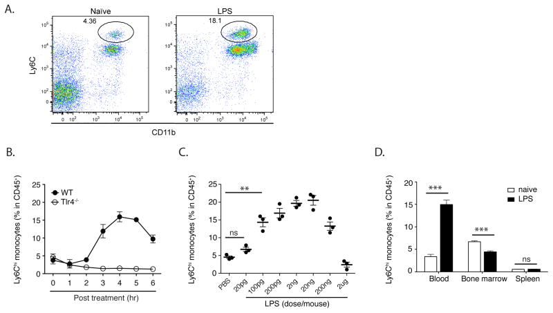
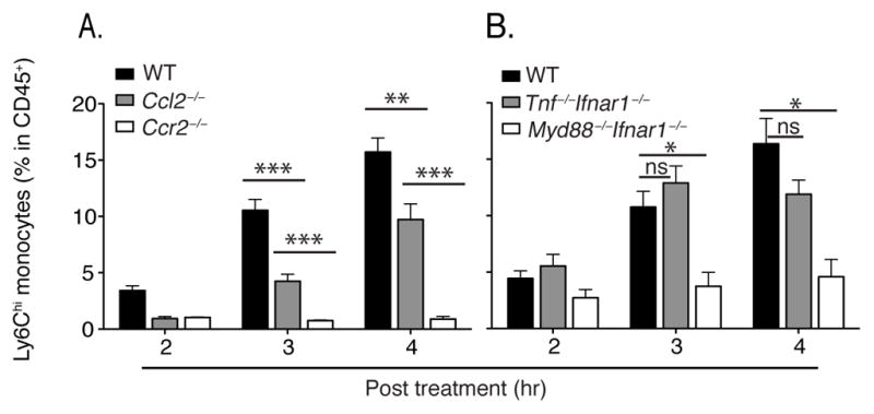
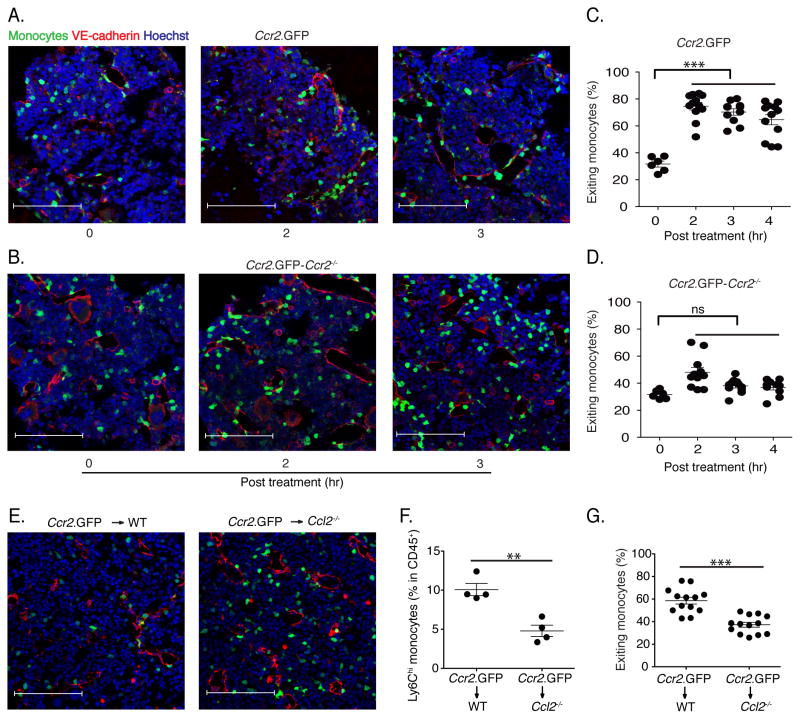
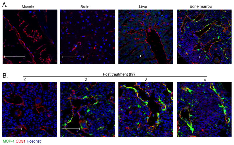
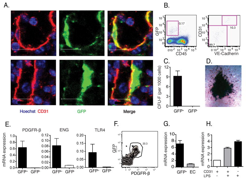
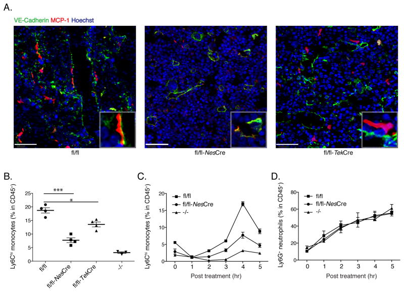
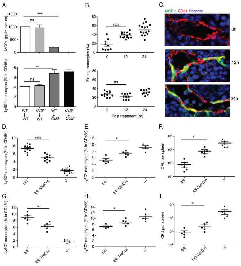
Inflammatory (Ly6C(hi) CCR2+) monocytes provide defense against infections but also contribute to autoimmune diseases and atherosclerosis. Monocytes originate from bone marrow and their entry into the bloodstream requires stimulation of CCR2 chemokine receptor by monocyte chemotactic protein-1 (MCP1). How monocyte emigration from bone marrow is triggered by remote infections remains unclear. We demonstrated that low concentrations of Toll-like receptor (TLR) ligands in the bloodstream drive CCR2-dependent emigration of monocytes from bone marrow. Bone marrow mesenchymal stem cells (MSCs) and their progeny, including CXC chemokine ligand (CXCL)12-abundant reticular (CAR) cells, rapidly expressed MCP1 in response to circulating TLR ligands or bacterial infection and induced monocyte trafficking into the bloodstream. Targeted deletion of MCP1 from MSCs impaired monocyte emigration from bone marrow. Our findings suggest that bone marrow MSCs and CAR cells respond to circulating microbial molecules and regulate bloodstream monocyte frequencies by secreting MCP1 in proximity to bone marrow vascular sinuses.


Figures

Fig. 1
Fig. 1. Low amounts of LPS drive monocyte emigration from bone marrow
(A) Increased circulating monocytes were identified as CD11b+Ly6Chi by flow cytometry. Flow cytometry plots are gated on CD45+ nucleated cells. (B) Wildtype (WT) and TLR4-deficient mice were injected intraperitoneally (i.p.) with 2ng LPS. Blood was obtained at the indicated time points following inoculation and analyzed by flow cytometry to determine CD11b+Ly6Chi monocyte percentages in CD45+ nucleated cells in the circulation. (C) WT mice were inoculated with PBS or 20pg to 2ug LPS and blood monocyte frequencies were determined 4 hr after LPS injection. (D) WT mice were inoculated with 2ng LPS. 4 hr later, blood, bone marrow and spleen monocyte frequencies were determined. Data are representative of 3–7 mice per group from at least three independent experiments, represented as mean +/− SEM.
Fig. 2
Fig. 2. LPS-induced emigration is CCR2, MCP1 and MyD88 dependent but TNF and type I interferon independent
(A, B) WT, Ccl2−/−, Ccr2−/−, Tnf−/−Ifnar1−/− and Myd88−/−Ifnar1−/− mice were inoculated with 20ng LPS. Blood monocyte frequencies were determined at indicated time points. Data are represented as mean +/− SEM.
Fig. 3
Fig. 3. Monocyte emigration is CCR2-dependent and is mediated by MCP1 produced by non-hematopoietic cells
(A and B) Fixed-frozen bone marrow sections from naïve and LPS-treated (2ng/mouse) CCR2 reporter mice (A) and CCR2 reporter-Ccr2−/− mice (B) were stained with Goat anti-mouse VE-cadherin and counterstained with Hoechst (63X). (C) Quantification of exiting monocyte percentage from CCR2 reporter and CCR2 reporter-Ccr2−/− mice following LPS treatment. Exiting monocyte percentages were calculated by dividing the total number of total GFP+ cells by the number of GFP+ cells within one cell distance of VE-cadherin+ endothelium. (E) Ccr2.GFP>WT and Ccr2.GFP>Ccl2−/− bone marrow chimeric mice were treated with 2ng/mouse LPS for 4hr. Fixed-frozen femurs were stained with anti-VE-cadherin. (F) Circulating monocyte frequencies in blood were determined in bone marrow chimeric mice. (G) The percentage of monocytes associated with VE-cadherin expessing endothelials, defined as exiting monocytes, in bone marrow was calculated. Data are representative of three independent experiments. Error bars indicate SEM. Scale bars indicate 90μm.
Fig. 4
Fig. 4. MCP1-producing cells in bone marrow co-localize with vascular sinuses
(A) WT>M1R bone marrow chimeric mice were inoculated with 20ng LPS and 4 hr later muscle, brain, liver and bone marrow were isolated, fixed and frozen and stained for CD31 expression and examined by confocal microscopy for GFP expression. Scale bars indicate 90μm. (B) Fixed-frozen bone marrow samples at the indicated time points following 20ng LPS inoculation were stained for CD31 expression and examined by confocal microscopy. Scale bars indicate 47μm. Data are representative of at least three independent experiments.
Fig. 5
Fig. 5. Marrow endothelial cells and tightly associated MSCs produce MCP1 and promote monocyte emigration
(A) M1R mice were inoculated with 20ng LPS and 4 hr later bone marrow was sectioned and stained. GFP+ MCP1 producing cells after LPS stimulation overlap (upper) or are tightly associated (lower) with CD31+ endothelial cells. Scale bars indicate 12μm. (B) Bone marrow from stimulated mice was harvested, dissociated and CD45GFP+ cells (left panel) were stained for expression of CD31 and VE-cadherin (right panel). (C) CD45GFP+ and CD45GFP cells were flow cytometrically-sorted from M1R mice following LPS inoculation and cultured for CFU-F. (D) CD45GFP+ cells were further differentiated in vitro to osteoblasts. (E) Pdgfrβ, Eng and Tlr4 expression in sorted cells were measured by Q-PCR. (F) Expression of GFP and PDGFRβ by CD45Ter119CD31Sca1 population in the bone marrow. (G) CD45Ter119GFP+ cells and CD45Ter119 CD31+Sca1+ endothelial cells were sorted and CXCL12 mRNA expression was quantified by Q-PCR. (H) CD45CD31GFP+ and CD45CD31+GFP+ cells were sorted and MCP1 mRNA expression was quantified by Q-PCR. Data are represented as mean +/− SEM.
Fig. 6
Fig. 6. MCP1 produced by Nestin-expressing MSCs is required for optimal monocyte emigration after LPS stimulation
Ccl2-RFPflox/flox, Ccl2-RFPflox/flox expressing Nes-Cre or Tek-Cre, and Ccl2−/− were inoculated with LPS. 4 hr post stimulation, (A) bone marrow was sectioned and stained. Scale bars indicate 40μm. (B) blood was obtained analyzed by flow cytometry to determine CD11b+Ly6Chi monocyte frequencies in the circulation. The emigration kinetics of CD11b+Ly6Chi monocytes and CD11b+Ly6G+ neutrophils are shown in (C) and (D). Error bars indicate SEM.
Fig. 7
Fig. 7. MCP1 production by bone marrow MSCs enhances resistance to Listeria monocytogenes infection
(A) Bone marrow chimeric mice were infected with 3000 L. monocytogenes. 24 hr after infection, serum was harvested for MCP1 ELISA (upper panel) and bone marrow cells were harvested to quantify Ly6Chi monocytes (lower panel). (B) CCR2 reporter (upper panel) and CCR2 reporter-Ccr2−/− mice (lower panel) were infected with L. monocytogenes. The percentages of exiting monocytes were quantified at different time points following infection. (C) WT-M1R bone marrow chimeric mice were infected with L. monocytogenes. Fixed-frozen sections were prepared at different time points and stained for CD31 expression, and examined by confocal microscopy. Data are representative of at least three independent experiments. Ccl2-RFPflox/flox, Ccl2-RFPflox/flox expressing Nes-Cre or Tek-Cre, and Ccl2−/− mice were infected with L. monocytogenes. CD11b+Ly6Chi monocyte frequencies in the circulation (D, G) and the bone marrow (E, H) were determined by flow cytometry at d1 post infection and the number of viable L. monocytogenes the spleens was quantified at d3 post infection (F, I). Data are represented as mean +/− SEM.

Comment in

References

    1. Aldridge JR, Jr, Moseley CE, Boltz DA, Negovetich NJ, Reynolds C, Franks J, Brown SA, Doherty PC, Webster RG, Thomas PG. TNF/iNOS-producing dendritic cells are the necessary evil of lethal influenza virus infection. Proc Natl Acad Sci U S A. 2009;106:5306–5311. - PMC - PubMed
    1. Andonegui G, Bonder CS, Green F, Mullaly SC, Zbytnuik L, Raharjo E, Kubes P. Endothelium-derived Toll-like receptor-4 is the key molecule in LPS-induced neutrophil sequestration into lungs. J Clin Invest. 2003;111:1011–1020. - PMC - PubMed
    1. Andonegui G, Zhou H, Bullard D, Kelly MM, Mullaly SC, McDonald B, Long EM, Robbins SM, Kubes P. Mice that exclusively express TLR4 on endothelial cells can efficiently clear a lethal systemic Gram-negative bacterial infection. J Clin Invest. 2009;119:1921–1930. - PMC - PubMed
    1. Boring L, Gosling J, Cleary M, Charo IF. Decreased lesion formation in CCR2−/− mice reveals a role for chemokines in the initiation of atherosclerosis. Nature. 1998;394:894–897. - PubMed
    1. Brenchley JM, Price DA, Schacker TW, Asher TE, Silvestri G, Rao S, Kazzaz Z, Bornstein E, Lambotte O, Altmann D, et al. Microbial translocation is a cause of systemic immune activation in chronic HIV infection. Nat Med. 2006;12:1365–1371. - PubMed

Publication types