. 2011 Apr 22;42(2):250-60.
doi: 10.1016/j.molcel.2011.03.010. Epub 2011 Mar 31.

A de novo protein binding pair by computational design and directed evolution

Affiliations

A de novo protein binding pair by computational design and directed evolution

John Karanicolas et al. Mol Cell. .

Abstract

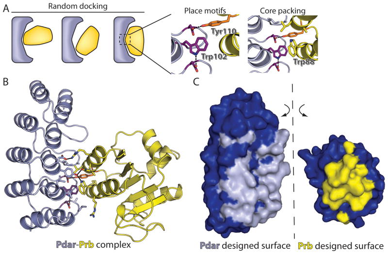
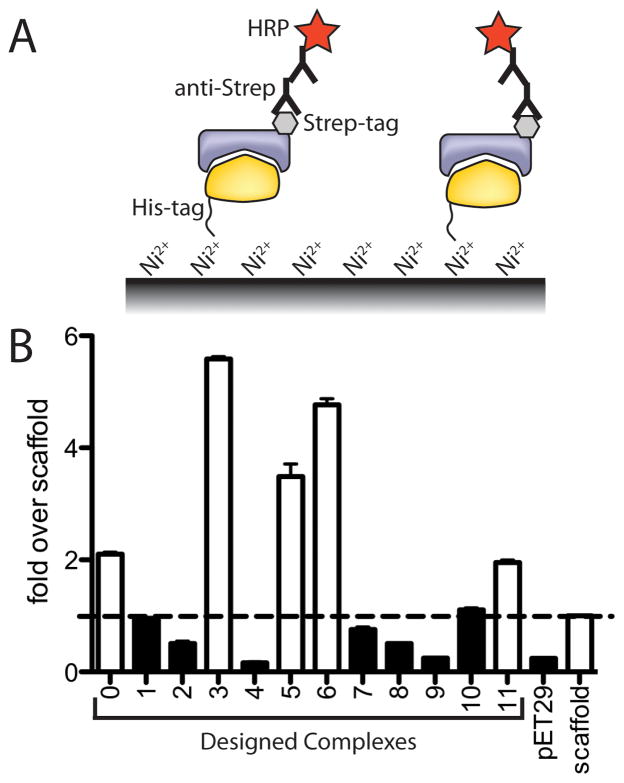
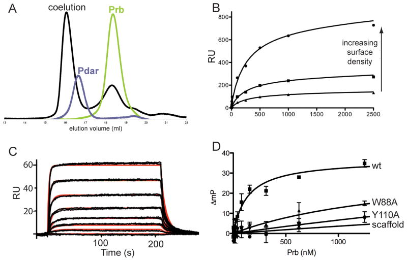
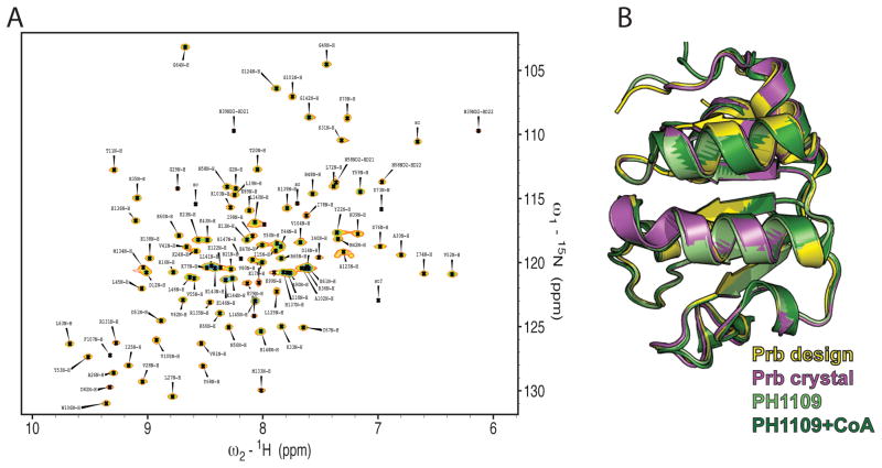
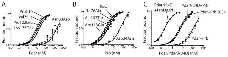
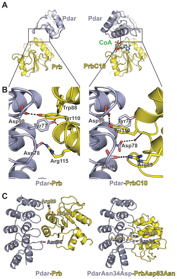
The de novo design of protein-protein interfaces is a stringent test of our understanding of the principles underlying protein-protein interactions and would enable unique approaches to biological and medical challenges. Here we describe a motif-based method to computationally design protein-protein complexes with native-like interface composition and interaction density. Using this method we designed a pair of proteins, Prb and Pdar, that heterodimerize with a Kd of 130 nM, 1000-fold tighter than any previously designed de novo protein-protein complex. Directed evolution identified two point mutations that improve affinity to 180 pM. Crystal structures of an affinity-matured complex reveal binding is entirely through the designed interface residues. Surprisingly, in the in vitro evolved complex one of the partners is rotated 180° relative to the original design model, yet still maintains the central computationally designed hotspot interaction and preserves the character of many peripheral interactions. This work demonstrates that high-affinity protein interfaces can be created by designing complementary interaction surfaces on two noninteracting partners and underscores remaining challenges.


Figures

Figure 1
Figure 1. Computational Design Approach
The Ankyrin Repeat protein (redesigned to become Pdar) is colored gray and its partner protein (redesigned from PH1109 to become Prb) is colored yellow. (A) The surfaces of each protein are first matched by general shape complementarity and local docking. Promising rigid body orientations are used in an attempt to place a central hydrogen-bonding tyrosine or tryptophan motif, followed by local design to enforce hydrophobic packing around the motif. (B) Overview of interfacial amino acids in the designed complex of Prb-Pdar. The central motifs are colored as in A. (C) Open-book surface view of the completely designed Prb-Pdar interface. Non-designed residues are colored blue, and designed interfacial residues are colored either gray (Pdar) or yellow (Prb). All figures containing molecular graphics were generated using PyMOL (DeLano, 2004).
Figure 2
Figure 2. Experimental screening for interacting computationally designed complexes
A) An ELISA screen for stably-associating designed protein complexes. Both partners, one His6-tagged and the other Strep-tagged, were co-expressed in E. coli. Complexes were then captured on a Nickel-derivatized surface detected with an anti-Strep antibody. B) Nearly half of the computationally designed protein-protein complexes (open bars) displayed signal more than two-fold over a control that replaces one side of Complex #11 with the wild-type protein used as a scaffold for the design (arbitrary identifiers 0–11 were assigned to the 12 designed pairs remaining after computational filtering).
Figure 3
Figure 3. Prb and Pdar form a hotspot-based high affinity protein complex with kinetics similar to those observed in natural protein assemblies
A) Prb and Pdar stably associate with one another to co-elute from a gel filtration column. B) Equilibrium surface plasmon resonance (SPR) measurements indicate that Prb and Pdar associate with Kd 60–90 nM. C) Kinetic SPR measurements at low surface density indicate that Prb and Pdar associate with kon of 7–9×10⁵ M−1 s−1 and koff of 0.05–0.1 s−1, which is in the range of natural protein-protein complexes and corresponds to Kd approximately 50–150 nM. Duplicate measurements are shown in black, and a 1:1 Langmuir fit is shown in red. D) Fluorescence polarization confirms that Prb and Pdar bind one another with a Kd of 135 nM. Furthermore, Pdar does not associate with wild-type PH1109, the protein scaffold used as a starting point in the Prb design. Mutation of key designed interfacial residues abrogates complex formation.
Figure 4
Figure 4. Interfacial residues of apo Prb can adopt the designed conformation, but may also be mobile
A) 2D 1H-15N HSQC NMR spectrum of Prb. Peaks are labeled with their respective sequential resonance assignments using the one-letter code of amino acids and the amino acid sequence number. Approximately thirty residues located at the designed interfacial positions are missing from the spectrum, suggesting that they may be conformationally dynamic. B) A crystal structure of the Prb protein (magenta) agrees well with both the design model (yellow, 0.54Å all-atom rmsd) and structures of the PH1109 scaffold either apo (light green, 0.41Å all-atom rmsd) or bound to CoA (dark green, 0.47Å all-atom rmsd).
Figure 5
Figure 5. Directed evolution identifies two point mutants that increase the affinity of Prb and Pdar by 720-fold
A) Reversion of each mutation within the affinity matured PrbC10 clone to its Prb identity indicates that Asn83Asp is responsible for most of PrbC10’s increase in affinity. B) Reversion of each mutation within the affinity matured PdarC1 clone to its Pdar identity indicates that Asn34Asp is responsible for most of PdarC1’s increase in affinity. C) The complex of Prb(Asn83Asp) and Pdar(Asp34Asn) binds with Kd of 180 pM, an increase in affinity of approximately 720-fold through only two mutations.
Figure 6
Figure 6. Structural characterization of Prb and a complex evolved from Prb-Pdar
A) The crystal structure of Prb bound to evolved clone 10 (PrbC10, yellow) bound to coenzyme A (green) and Pdar (gray, right) is rotated 180 degrees relative to the designed Prb (yellow) bound to Pdar (gray, left). B) The evolved interface makes several interactions symmetric to those designed for Prb-Pdar. Pdar is shown in equivalent orientations, highlighting that the complex in the crystal structure is rotated 180 degrees relative to the design model, such that a Prb(Tyr110)/Pdar(Asp98) hydrogen bond is maintained. This generates several similar interactions, such as the Prb(Arg115)/Pdar(Asp78) and PrbC10(Arg89)/Pdar(Asp65) salt bridges. C) The rotated orientation places PrbC10(Arg89) adjacent to Pdar(Asn34), which is mutated to an aspartate in several affinity-matured Pdar variants. Prb(Asp83), which is mutated to asparagine in PrbC10 is brought towards the center of the interface in the rotated orientation. The model of Pdar(Asn34Asp) paired with Prb(Asp83Asn) was generated by computationally mutating relevant amino acids within the Pdar-PrbC10 crystal structure, followed by repacking and energy minimization.

References

    1. Aharoni A, Gaidukov L, Khersonsky O, Mc QGS, Roodveldt C, Tawfik DS. The ‘evolvability’ of promiscuous protein functions. Nat Genet. 2005;37:73–76. - PubMed
    1. Aloy P, Ceulemans H, Stark A, Russell RB. The relationship between sequence and interaction divergence in proteins. J Mol Biol. 2003;332:989–998. - PubMed
    1. Apgar JR, Gutwin KN, Keating AE. Predicting helix orientation for coiled-coil dimers. Proteins. 2008;72:1048–1065. - PMC - PubMed
    1. Batchelor AH, Piper DE, de la Brousse FC, McKnight SL, Wolberger C. The structure of GABPalpha/beta: an ETS domain- ankyrin repeat heterodimer bound to DNA. Science. 1998;279:1037–1041. - PubMed
    1. Binz HK, Amstutz P, Pluckthun A. Engineering novel binding proteins from nonimmunoglobulin domains. Nat Biotechnol. 2005;23:1257–1268. - PubMed

Publication types