

Review
doi: 10.1016/j.micinf.2011.03.002.
Epub 2011 Mar 31.
Lipids, apoptosis, and cross-presentation: links in the chain of host defense against Mycobacterium tuberculosis
Affiliations
- PMID: 21458584
- PMCID: PMC3130819
- DOI: 10.1016/j.micinf.2011.03.002
Item in Clipboard
Review
Lipids, apoptosis, and cross-presentation: links in the chain of host defense against Mycobacterium tuberculosis
Microbes Infect.
2011 Aug.
Abstract
Eicosanoids regulate whether human and murine macrophages infected with Mycobacterium tuberculosis die by apoptosis or necrosis. The death modality is important since apoptosis is associated with diminished pathogen viability and should be viewed as a form of innate immunity. Apoptotic vesicles derived from infected macrophages are also an important source of bacterial antigens that can be acquired by dendritic cells to prime antigen-specific T cells. This review integrates in vitro and in vivo data on how apoptosis of infected macrophages is linked to development of T cell immunity against M. tuberculosis.
Copyright © 2011 Institut Pasteur. Published by Elsevier SAS. All rights reserved.
Figures
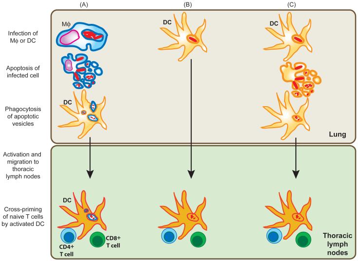
Three models of how DC could acquire mycobacterial antigens during Mtb infection of the lung and prime naïve Mtb-specific T cells in the draining lymph nodes (LN) of the infected host. In the first model (A), infected macrophages in the lung undergoing apoptosis would generate apoptotic vesicles containing mycobacterial antigens. Pulmonary DC could take up these blebs and vesicles and migrate to the draining LN. Once there, the DC would be competent to prime naïve T cells. Antigen in the endocytic pathway could enter the class II MHC pathway leading to priming of CD4+ T cells. Cross-presentation of these antigens by the class I MHC pathway could prime CD8+ T cells. Alternately (B), infected DC could migrate from the lung to the draining LN, where the infected DC could directly prime CD4+ and CD8+ T cells. In the third model, Mtb infected DC may also undergo apoptosis in the lung. Analogous to (A), uninfected DC could engulf the apoptotic vesicles derived from infected DC, migrate to the LN and prime naïve T cells. Not shown here is another permutation in which infected DC or macrophages (B) could migrate to the draining LN, undergo apoptosis, and the mycobacterial antigen laden apoptotic vesicles taken up by LN resident DC. The antigens acquired in this manner by the LN-resident DC could then lead to the priming of naïve T cells.

Macrophages are briefly infected in vitro with Mtb, washed and instilled into the lungs of recipient mice. While in the lung the transferred macrophage may undergo Mtb induced cell death. For example, macrophages lacking the enzyme 5-lipoxygenase (5-LO) are prone to die by apoptosis after infection with virulent Mtb. During apoptosis the plasma membrane remains intact as the nucleus fragments and cellular components along with bacterial debris and Mtb-containing phagosomes are contained in apoptotic blebs and vesicles. Apoptosis of infected macrophages is correlated with innate control of bacterial replication alhtough the mechanism remains unclear. In addition, DC that engulf apoptotic vesicles that contain Mtb or Mtb products can cross-present the bacterial antigens to CD8+ T cells. In contrast, necrosis, which is characterized by the disintegration of the plasma membrane, swelling of the cell and organelle shrinkage and rupture, allows for the bacteria to escape into the extracellular space and infect surrounding macrophages. Mtb-infected macrophages that lack the enzyme prostaglandin E synthase (PGES) are prone to necrosis.
References
-
- Godfrey DI, Rossjohn J, McCluskey J. The fidelity, occasional promiscuity, and versatility of T cell receptor recognition. Immunity. 2008;28:304–314. - PubMed
-
- Cresswell P, Ackerman AL, Giodini A, Peaper DR, Wearsch PA. Mechanisms of MHC class I-restricted antigen processing and cross-presentation. Immunol Rev. 2005;207:145–157. - PubMed
-
- Hume DA. Applications of myeloid-specific promoters in transgenic mice support in vivo imaging and functional genomics but do not support the concept of distinct macrophage and dendritic cell lineages or roles in immunity. Journal of Leukocyte Biology. 2010 - PubMed
-
- Hume DA. Macrophages as APC and the dendritic cell myth. J Immunol. 2008;181:5829–5835. - PubMed
Publication types
MeSH terms
Substances
Grants and funding
LinkOut - more resources
Full Text Sources
Medical
