. 2011;269(1):38-45.
doi: 10.1016/j.cellimm.2011.03.008. Epub 2011 Mar 15.

The oral commensal, Streptococcus gordonii, synergizes with Tat protein to induce HIV-1 promoter activation in monocytes/macrophages

Affiliations

The oral commensal, Streptococcus gordonii, synergizes with Tat protein to induce HIV-1 promoter activation in monocytes/macrophages

Octavio A González et al. Cell Immunol. 2011.

Abstract

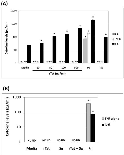
Trans-activator of transcription (Tat) is an HIV-1 protein essential for viral replication. Oral periodontopathogens (e.g. Fusobacterium nucleatum) enhance HIV-1LTR promoter activation in monocytes/macrophages in absence of Tat; however, some oral commensals fail to trigger this response. We sought to determine the effect of Tat on HIV-1LTR promoter activation induced by the representative oral commensal Streptococcus gordonii in monocytes/macrophages. S. gordonii enhanced HIV-1LTR reactivation in THP89GFP (Tat(+)), but not in BF24 (Tat(-)) cells. Interestingly, S. gordonii, but not Streptococcus sanguinis enhanced HIV-1LTR activation in the presence of recombinant Tat in BF24 cells. This response correlated with IL-8 but not TNFα or IL-6 production, and was abrogated by the NFκB inhibitor BAY 11-7082. Kinetics of NFκB-RelA activation did not explain the S. gordonii-induced HIV-1LTR activation in presence of Tat. These results suggest that S. gordonii-induced HIV-1 reactivation in monocytes/macrophages is Tat-dependent and appears to involve NFκB activation.


Figures

FIGURE 1
FIGURE 1. S. gordonii enhances HIV-1LTR activation in THP89GFP, but not in BF24 monocytes/macrophages
2.5×10⁵ cells/ml were challenged with 10 μg/ml S. gordonii extract and (A) HIV-1/CAT activity in BF24 cell lysates was determined by CAT-ELISA, and (B) HIV-1/EGFP activity as well as (C) p24 levels were determined in THP89GFP cells by fluorometry and ELISA respectively at different time points until 72h as described in methods. BF24 cells challenged with 10 μg/ml of P. gingivalis extract or incubated only with media (Mock) were used as positive and negative controls respectively for HIV-1/CAT activation. The figures are representative of at least 2 independent experiments by triplicate (n=6). Data are expressed as means ± standard deviations. The asterisk (*) denotes P ≤ 0.01 compared to the results for the control (mock), as determined by a Kruskal Wallis ANOVA with Dunnett’s test for multiple comparisons. ND = Not detected.
FIGURE 2
FIGURE 2. Oral commensals S. gordonii and S. sanguinis fail to induce HIV-1LTR promoter activation in B24 monocytes/macrophages
BF24 cells (2.5 × 10⁵/well) were seeded into 24-well plates with 1 ml of media and left either untreated or challenged with different concentrations of bacterial extracts from S. gordonii and S. sanguinis for 16h. (A) IL-8 levels in supernatants and (B) HIV-1/CAT activity in cell lysates were determined by ELISA as described in methods. BF24 cells exposed to sodium butyrate (NB) or incubated only with media (Mock) were used as positive and negative controls respectively for HIV-1 promoter activity. The figures are representative of at least 2 independent experiments in triplicate (n=6). Data are expressed as means ± standard deviations.*, p ≤ 0.01 compared to the results for the control (mock), as determined by a Mann-Whitney U test on ranks. ND= Not detected.
FIGURE 3
FIGURE 3. S. gordonii synergizes with rTat to enhance HIV-1LTR promoter activation and IL-8 production
BF24 cells (2.5×10⁵/well) were first incubated only with media or in presence of several concentrations of rTat (10-500 ng/ml). (A) IL-8 levels in supernatants and (B) HIV-1/CAT promoter activity in cell lysates were determined 16h post-treatment as described in methods. Using the same cell density, BF24 cells were pre-incubated 30 minutes with either 100 ng/ml rTat or media followed by challenge with different concentrations of bacterial extracts from S. gordonii or S. sanguinis and HIV-1/CAT promoter activity in cell lysates (C and D) or IL-8 production in supernatants (E and F) were determined by ELISA as described in methods. The figures are representative of at least 2 independent experiments by triplicate (n=6). Data are expressed as means ± standard deviations. Values that were significantly different (p≤0.01) from mock/media (*) or with/without rTat (#) as determined by a Kruskal Wallis ANOVA with Dunnett’s test for multiple comparisons (A and B) or a Mann-Whitney U test on ranks (C-F). ND= Not detected.
FIGURE 4
FIGURE 4. Pro-inflammatory cytokine production by B24 monocytes/macrophages in response to S. gordonii and rTat
BF24 cells (2.5 × 10⁵/well) were seeded in 24-well plates with 1 ml of media and cytokine levels were determined by ELISA in supernatants 16h after different treatments as described in methods. (A) Levels of IL-6, TNFα and IL-8 in supernatants from BF24 cells challenged with different concentrations of either rTat (10-500 ng/ml) or 10 μg/ml of S. gordonii (Sg) extract are shown. (B) Levels of pro-inflammatory cytokines TNFα and IL-6 induced by either 10 μg/ml of S. gordonii extract or 100 ng/ml of rTat individually or in combination were determined by ELISA. BF24 cells exposed to oral Gram-negative P. gingivalis (Pg) or F. nucleatum (Fn) extracts and cells incubated only with media were used as positive and negative controls respectively for cytokine production. The figures are representative of at least 2 independent experiments in triplicate (n=6). Data are expressed as means ± standard deviations.*, p ≤ 0.01 compared to the results for the control (mock/media), as determined by a Kruskal Wallis ANOVA with Dunnett’s test for multiple comparisons (rTat) or a Mann-Whitney U test ranks (Pg and Sg vs. media and Fn vs. media). ND= Not detected.
FIGURE 5
FIGURE 5. NFκB activation is involved in HIV-1LTR promoter activation induced by S. gordonii in presence of rTat
BF24 cells (2.5×10⁵/ml) pre-incubated 30 minutes 100 ng/ml rTat were challenged with 10 μg/ml of bacterial extract from S. gordonii either in presence or absence of different concentrations of NFκB inhibitor BAY 11-7082 for 16h and (A) HIV-1/CAT promoter activity in cell lysates, or (B) IL-8 levels in supernatants were determined by ELISA as described in methods. BF24 cells incubated only with media (mock) were used as a negative control (C) Kinetics of NFκB-RelA(p65) activation was evaluated in nuclear extracts from BF24 cells (2.5×10⁵/ml) challenged with 10 μg/ml of bacterial extract from S. gordonii, 100 ng/ml rTat, or both (Sg + rTat), and 10 μg/ml F. nucleatum extract by TransAM as described in methods for the indicated times. The figures are representative of at least 2 independent experiments by triplicate (n=6). Data are expressed as means ± standard deviations. Values that were significantly different (p≤0.01) comparing presence of NFκB inhibitor BAY 11-7082 for HIV-1/CAT and IL-8 determined by a Mann-Whitney U test on ranks (A and B) are indicated by asterisks (*), or by Kruskal Wallis ANOVA with Dunnett’s test for multiple comparisons (C) For NFκB/p65 levels the asterisks (*) denote significantly different (p≤0.01) from S. gordonii alone and pound sign (#) denotes significantly different (p≤0.01) from S. gordonii/F. nucleatum induced NFκB/p65 levels at 0 time.

Similar articles

Cited by

References

    1. Alexaki A, Liu Y, Wigdahl B. Cellular reservoirs of HIV-1 and their role in viral persistence. Curr HIV Res. 2008;6:388–400. - PMC - PubMed
    1. Coleman CM, Wu L. HIV interactions with monocytes and dendritic cells: viral latency and reservoirs. Retrovirology. 2009;6:51. - PMC - PubMed
    1. Richman DD, Margolis DM, Delaney M, Greene WC, Hazuda D, Pomerantz RJ. The challenge of finding a cure for HIV infection. Science. 2009;323:1304–1307. - PubMed
    1. Pereira LA, Bentley K, Peeters A, Churchill MJ, Deacon NJ. A compilation of cellular transcription factor interactions with the HIV-1 LTR promoter. Nucleic Acids Res. 2000;28:663–668. - PMC - PubMed
    1. Rohr O, Marban C, Aunis D, Schaeffer E. Regulation of HIV-1 gene transcription: from lymphocytes to microglial cells. J Leukoc Biol. 2003;74:736–749. - PubMed

Publication types

MeSH terms

Substances

LinkOut - more resources