Comparative Study
. 2011 Jun;32(6):935-43.
doi: 10.1093/carcin/bgr059. Epub 2011 Mar 31.

Slit-2 facilitates interaction of P-cadherin with Robo-3 and inhibits cell migration in an oral squamous cell carcinoma cell line

Affiliations
Comparative Study

Slit-2 facilitates interaction of P-cadherin with Robo-3 and inhibits cell migration in an oral squamous cell carcinoma cell line

Karin Bauer et al. Carcinogenesis. 2011 Jun.

Abstract

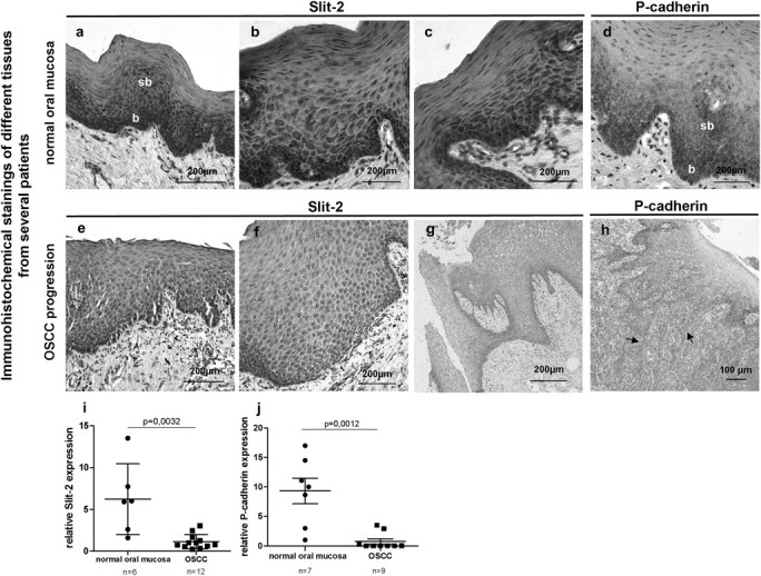
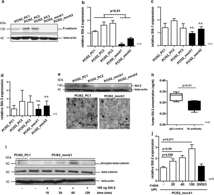
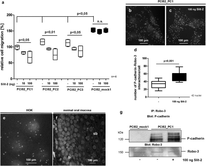
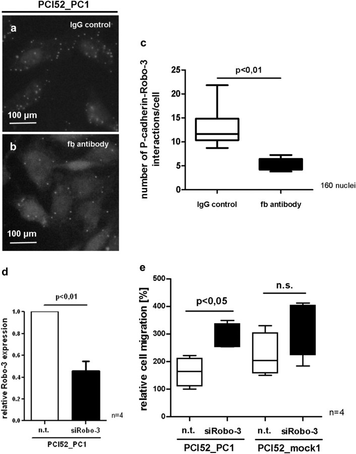
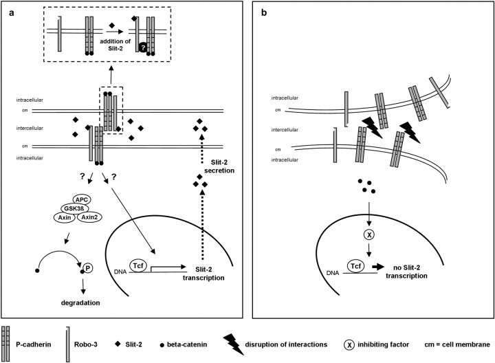
Slits are a group of secreted glycoproteins that act as molecular guidance cues in cellular migration. Recently, several studies demonstrated that Slit-2 can operate as candidate tumour suppressor protein in various tissues. In this study, we show Slit-2 expression in basal cell layers of normal oral mucosa colocalized with P-cadherin expression. In contrast, there is a loss of Slit-2 and P-cadherin expression in mucosa of oral squamous cell carcinoma (OSCC). Our in vitro investigations reveal a correlation of P-cadherin and Slit-2 expression: OSCC cells with induced P-cadherin expression (PCI52_PC) display an increased Slit-2 expression. However, abrogating P-cadherin function with a function-blocking antibody decreases Slit-2 secretion confirming a direct link between P-cadherin and Slit-2. Moreover, experiments with OSCC cells show that Slit-2 interferes with a Wnt related signalling pathway, which in turn affects Slit-2 expression in a feedback loop. Functionally, transwell migration assays demonstrate a Slit-2 dose-dependent decrease of PCI52_PC cell migration. However, there is no influence on migration in mock control cells. Responsible for this migration block might be an interaction of P-cadherin with Roundabout (Robo)-3, a high affinity receptor of Slit-2. Indeed, proximity ligation assays exhibit P-cadherin/Robo-3 interactions on PCI52_PC cells. Additionally, we detect a modulation of this interaction by addition of recombinant Slit-2. Down-regulation of Robo-3 expression via small interfering RNA neutralizes Slit-2 induced migration block in PCI52_PC cells. In summary, our experiments show antitumorigenic effects of Slit-2 on P-cadherin expressing OSCC cells supposedly via modulation of Robo-3 interaction.


Figures

Fig. 1.
Fig. 1.
Representative immunohistochemical stainings of different tissues from several patients show that in normal oral mucosa, Slit-2 expression (ac) is localized in basal and suprabasal epithelial cell layers like P-cadherin (d). During neoplastic transformation of OSCC, Slit-2 reveals a rather low and dislocalized expression pattern in the oral mucosa (eg). Tissue sections of OSCC patients display a loss of membrane-bound P-cadherin at the invasion front of OSCC (h). qRT–PCR of cDNA obtained from snap frozen tissues confirms a significant decrease of Slit-2 (i) and P-cadherin (j) expression in OSCC patients compared with healthy controls (b, basal; sb, suprabasal cell layers of normal oral mucosa; arrows, invasion front).
Fig. 2.
Fig. 2.
Western blot analysis displays a strong P-cadherin expression in the stably transfected OSCC cell line PCI 52 (PCI52_PC1-3), whereas no P-cadherin expression can be seen in mock transfected OSCC cells (PCI52_mock1-2) (a). After P-cadherin overexpression Slit-2 expression increases significantly in PCI52_PC cells compared with PCI52_mock cells (b), whereas both Slit-1 and Slit-3 expression reveal no correlation with P-cadherin expression (c and d). Western blot analysis shows a higher Slit-2 protein level in PCI52_PC cells than in PCI52_mock cells (e). Immunocytochemical stainings affirm a stronger Slit-2 expression in PCI52_PC cells contact each other (f, see arrows) in contrast to sparse Slit-2 expression in a few PCI52_mock single cells (g, see arrows). Blocking P-cadherin function with function blocking (fb) P-cadherin antibody results in a significant decrease of Slit-2 secretion in PCI52_PC cells compared with cells with IgG control antibody treatment (h). Within an hour after Slit-2 incubation phosphorylation of beta-catenin appears in PCI52_mock cells, which have only trace amounts of phospho-beta-catenin (i). Inhibition of the beta-catenin/Lef/Tcf complex with FH535 inhibitor in PCI52_mock cells results in a dose-dependent increase of Slit-2 expression (j).
Fig. 3.
Fig. 3.
After addition of Slit-2 PCI52_PC cells display a dose-dependent decrease in cell migration, whereas PCI52_mock cells do not show any reaction on cell migration after Slit-2 addition (a). PLAs demonstrate an interaction of P-cadherin with Robo-3 receptor in vitro on PCI52_PC cells (b). After addition of Slit-2 to PCI52_PC cells, an increased number of P-cadherin/Robo-3 interactions can be observed (c and d). P-cadherin/Robo-3 interactions can also be seen in human oral keratinocytes (HOK) (e) and in basal cell layers of normal oral mucosa (f). Coimmunoprecipitation experiments confirm the occurrence of increased P-cadherin/Robo-3 interactions following Slit-2 treatment (g) (b = basal, sb = suprabasal cell layers of normal oral mucosa).
Fig. 4.
Fig. 4.
After blocking P-cadherin function with function blocking (fb) P-cadherin antibody, PCI52_PC cells reveal a significant decrease of P-cadherin/Robo-3 interactions by PLAs (ac). Knockdown of Robo-3 with siRNA (d) in PCI52_PC and PCI52_mock cells results in a significant increase in cell migration in Slit-2 producing PCI52_PC cells. However, in PCI52_mock cells, there is no significant migration effect after Robo-3 knockdown (e) (n.t. = non-targeting).
Fig. 5.
Fig. 5.
Hypothetical model of the formation of Slit-2 induced P-cadherin/Robo-3 complex and subsequent effects on Slit-2 transcription and secretion: Activation of Robo-3 by Slit-2 results in the formation of a complex between Robo-3 and P-cadherin mediated by still unknown molecules (a, small picture). Extracellular Slit-2 binds to Robo-3 receptor with subsequent abundant cytosolic beta-catenin phosphorylation and degradation. Consequently, further inhibiting transcription factors cannot be expressed thus supporting Slit-2 transcription in a feedback loop. Additionally, P-cadherin–P-cadherin interactions affect Slit-2 expression and facilitate Slit-2 secretion (a). In contrast, disruption of P-cadherin–P-cadherin interactions causes a diminished Slit-2 secretion and a decrease of P-cadherin/Robo-3 interactions. Because of the disrupted cell adhesion and decreased Slit-2 secretion, beta-catenin is not phosphorylated possibly due to GSK3-beta inactivation. Thus, beta-catenin is translocated to the nucleus where genes are activated which in turn might contribute to the inhibition of Slit-2 transcription (b).

References

    1. Bray F, et al. Estimates of cancer incidence and mortality in Europe in 1995. Eur. J. Cancer. 2002;38:99–166. - PubMed
    1. Takeichi M. Cadherins: a molecular family important in selective cell-cell adhesion. Annu. Rev. Biochem. 1990;59:237–252. - PubMed
    1. Drees F, et al. Alpha-catenin is a molecular switch that binds E-cadherin-beta-catenin and regulates actin-filament assembly. Cell. 2005;123:903–915. - PMC - PubMed
    1. Wheelock MJ, et al. Cadherin-mediated cellular signaling. Curr. Opin. Cell Biol. 2003;15:509–514. - PubMed
    1. Shimoyama Y, et al. Cadherin cell-adhesion molecules in human epithelial tissues and carcinomas. Cancer Res. 1989;49:2128–2133. - PubMed

Publication types

MeSH terms