

doi: 10.1101/pdb.prot5595.
Time-lapse imaging of postimplantation mouse embryos
- PMID: 21460044
- PMCID: PMC6800219
- DOI: 10.1101/pdb.prot5595
Item in Clipboard
Time-lapse imaging of postimplantation mouse embryos
Cold Spring Harb Protoc.
.
No abstract available
Figures
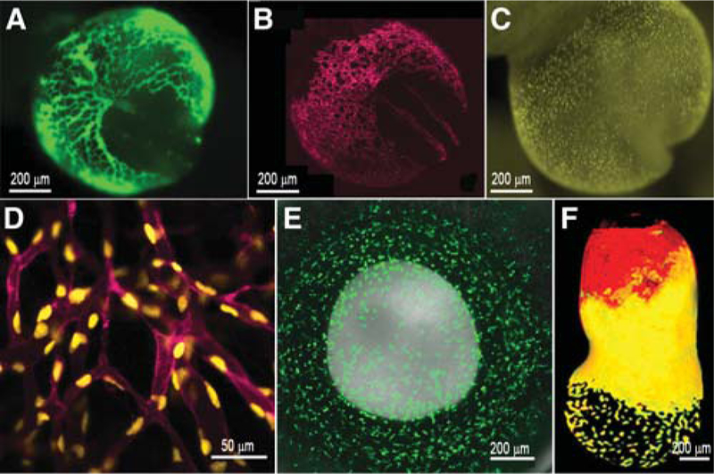
Fluorescent transgene expression in mouse embryos. (A) β-globin marks primitive erythroblasts in circulation within the yolk sac and embryo proper at 8.5 dpc. (B,C) The Flk1 promoter is used to drive membrane mCherry (B) and nuclear yellow fluorescent protein (YFP) (C) in endothelial cells of the developing embryo at 8.5 dpc. (D) A close-up image of vessels expressing YFP and mCherry in distinct subcellular localizations. (E) Green fluorescent protein (GFP) expression driven by the cfms promoter in the neonatal eye (postnatal day 1), marking macrophages. (F) Expression of a double transgenic embryo at 7.0 dpc expressing GFP and red fluorescent protein (RFP) in cells derived from the visceral endoderm. (F, Reprinted from Hadjantonakis et al. 2008 with permission from Elsevier © 2008.)

Example of a commercial heater box. Static culture system for time-lapse imaging of postimplantation mouse embryos. The microscope and static culture are housed in a temperature- (37°C) and CO2-regulated Plexiglas chamber. The gas mixture for the culture is humidified using a bubbler, and an inlet to the heated stage allows for ample gas exchange during static culture.
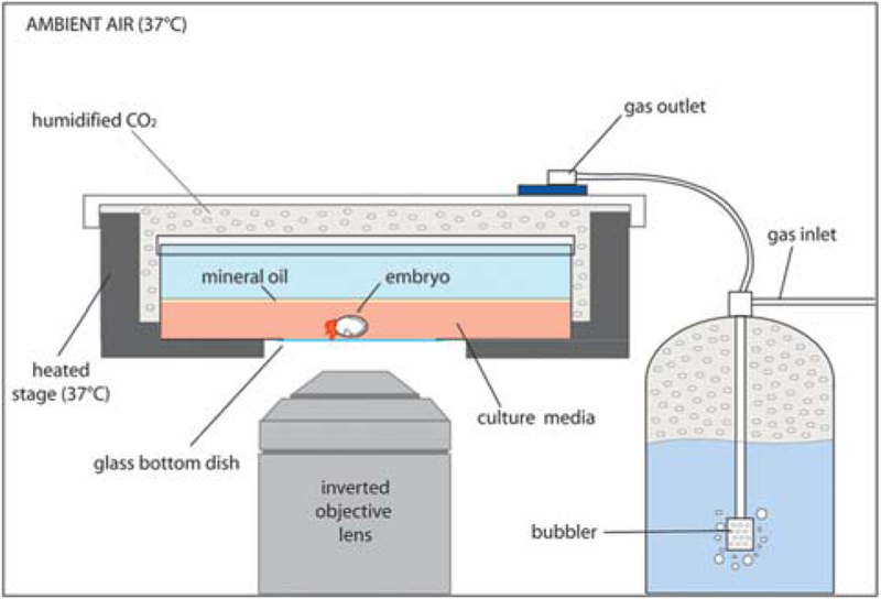
Schematic of heated culture chamber on an inverted microscope. The embryo is positioned inside a 37°C heated stage and covered with a gas outlet, allowing for humidified CO2 gas exchange within the chamber.
References
MeSH terms
Grants and funding
LinkOut - more resources
Full Text Sources