. 2011 Jun 17;286(24):21180-90.
doi: 10.1074/jbc.M110.203281. Epub 2011 Apr 1.

Glycomic analyses of mouse models of congenital muscular dystrophy

Affiliations

Glycomic analyses of mouse models of congenital muscular dystrophy

Stephanie H Stalnaker et al. J Biol Chem. .

Abstract

Dystroglycanopathies are a subset of congenital muscular dystrophies wherein α-dystroglycan (α-DG) is hypoglycosylated. α-DG is an extensively O-glycosylated extracellular matrix-binding protein and a key component of the dystrophin-glycoprotein complex. Previous studies have shown α-DG to be post-translationally modified by both O-GalNAc- and O-mannose-initiated glycan structures. Mutations in defined or putative glycosyltransferase genes involved in O-mannosylation are associated with a loss of ligand-binding activity of α-DG and are causal for various forms of congenital muscular dystrophy. In this study, we sought to perform glycomic analysis on brain O-linked glycan structures released from proteins of three different knock-out mouse models associated with O-mannosylation (POMGnT1, LARGE (Myd), and DAG1(-/-)). Using mass spectrometry approaches, we were able to identify nine O-mannose-initiated and 25 O-GalNAc-initiated glycan structures in wild-type littermate control mouse brains. Through our analysis, we were able to confirm that POMGnT1 is essential for the extension of all observed O-mannose glycan structures with β1,2-linked GlcNAc. Loss of LARGE expression in the Myd mouse had no observable effect on the O-mannose-initiated glycan structures characterized here. Interestingly, we also determined that similar amounts of O-mannose-initiated glycan structures are present on brain proteins from α-DG-lacking mice (DAG1) compared with wild-type mice, indicating that there must be additional proteins that are O-mannosylated in the mammalian brain. Our findings illustrate that classical β1,2-elongation and β1,6-GlcNAc branching of O-mannose glycan structures are dependent upon the POMGnT1 enzyme and that O-mannosylation is not limited solely to α-DG in the brain.


Figures

FIGURE 1.
FIGURE 1.
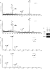
O-Glycans released from POMGnT1+/+ and POMGnT1−/− mouse brain proteins. a and b, O-glycans were released from protein powder made from POMGnT1+/+ and POMGnT1−/− mouse brain proteins by reductive β-elimination. Through comparison of the full MS scans of POMGnT1+/+ and POMGnT1−/−, we were able to observe the absence of prominent O-mannose structures in POMGnT1−/−. c and d, from the MS/MS scan (m/z 1256), the disialylated Tn antigen is unaffected in both POMGnT1+/+ and POMGnT1−/−. e, antibody IIH6, which recognizes the fully glycosylated, functionally active form of α-DG, shows absence of functionally active α-DG in POMGnT1−/− brains. WB, Western blot. f, individual monosaccharides were released from both POMGnT1+/+ and POMGnT1−/− mouse brain proteins, and the relative abundances of O-mannitol were compared. From the two data sets, proteins carrying non-extended O-mannose structures were determined to be enriched by ∼2.4-fold in the POMGnT1−/− animals. PAD, pulsed amperometric detection; Std, standard.
FIGURE 2.
FIGURE 2.
Linkage analysis of recombinant POMGnT1 by NMR. Two-dimensional 1H-13C correlation spectra show signals derived from the carbohydrate portion of the Ac-YVEP-(GlcNAc-β1,2-Man-α)-TAV-NH2 glycopeptide prepared from POMGnT1 action on the mannosylated peptide. A and B, sections from a heteronuclear multiple-quantum coherence spectrum, where the cross-peaks are at the coordinates of pairs of directly bonded protons and carbons. C and D, similar regions from the HMBC spectrum, where the peaks are at the coordinates of a proton and carbons two or three bonds away. The lines correlate peaks in the spectra that derive from the same protons associated with respective glycosidic linkages. Peaks in C show connections between the H-2 proton of Man and C-1 of GlcNAc, establishing the GlcNAc C-1 to Man C-2 glycosidic linkage, and additionally the linkage from the Thr-β proton to Man C-1. These are confirmed in the other direction across the linkages by the peaks in D.
FIGURE 3.
FIGURE 3.
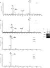
O-Glycans released from LARGE+/+ and LARGE−/− mouse brain proteins. a and b, O-glycans were released from protein powder made from LARGE+/+ and LARGE−/− mouse brain proteins. Through comparison of the full MS scans, we observed no difference in the prominent O-glycan structures that were detected in LARGE+/+ and LARGE−/− mouse brain proteins. c and d, MS/MS fragmentation spectra (m/z 1100) indicate the presence of the classical O-Man tetrasaccharide in both the LARGE+/+ and LARGE−/− mouse brains. e, antibody IIH6, which recognizes the fully glycosylated, functionally active form of α-DG, shows absence of functionally active α-DG in LARGE−/− brains. WB, Western blot.
FIGURE 4.
FIGURE 4.
O-Glycans released from α-DG+/+ and α-DG−/− (GFAP-Cre/DAG1) mouse brain proteins. a and b, O-glycans were released from protein powder of α-DG+/+ and α-DG−/− cerebrums. Through comparison of the full scan, we observed no difference in the amount of prominent O-mannose-initiated glycan structures detected in the α-DG+/+ and α-DG−/− proteins. c and d, MS/MS fragmentation spectra (m/z 912) indicate the presence of the fucosylated O-mannose trisaccharide structure in both the α-DG+/+ and α-DG−/− mouse brains. e, antibody IIH6, which recognizes the fully glycosylated, functionally active form of α-DG, indicates an order of magnitude decrease in the amount of α-DG levels present in the α-DG−/− brain compared with the α-DG+/+ brain. WB, Western blot.

References

    1. Lisi M. T., Cohn R. D. (2007) Biochim. Biophys. Acta 1772, 159–172 - PubMed
    1. Ibraghimov-Beskrovnaya O., Ervasti J. M., Leveille C. J., Slaughter C. A., Sernett S. W., Campbell K. P. (1992) Nature 355, 696–702 - PubMed
    1. Ervasti J. M., Campbell K. P. (1993) J. Cell Biol. 122, 809–823 - PMC - PubMed
    1. Barresi R., Campbell K. P. (2006) J. Cell Sci. 119, 199–207 - PubMed
    1. Martin P. T. (2007) Curr. Mol. Med. 7, 417–425 - PMC - PubMed

Publication types