. 2011 May;17(5):903-11.
doi: 10.1261/rna.2552811. Epub 2011 Apr 1.

Structural implications into dsRNA binding and RNA silencing suppression by NS3 protein of Rice Hoja Blanca Tenuivirus

Affiliations

Structural implications into dsRNA binding and RNA silencing suppression by NS3 protein of Rice Hoja Blanca Tenuivirus

Xia Yang et al. RNA. 2011 May.

Abstract

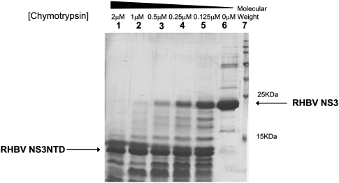
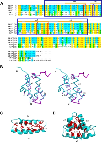
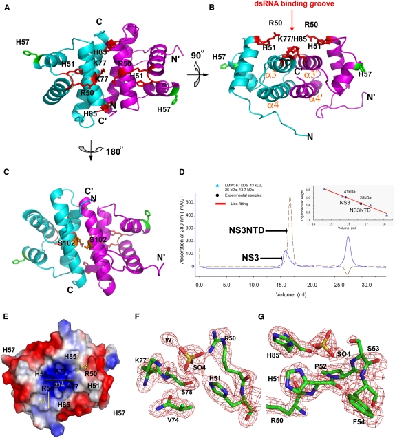
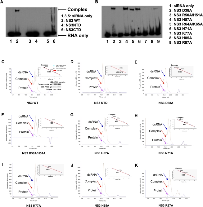
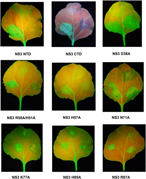
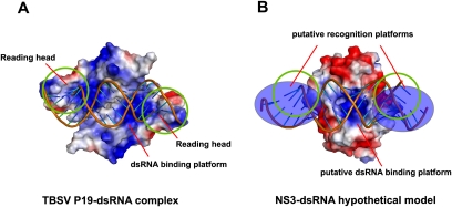
Rice Hoja Blanca Tenuivirus (RHBV), a negative strand RNA virus, has been identified to infect rice and is widely transmitted by the insect vector. NS3 protein encoded by RHBV RNA3 was reported to be a potent RNAi suppressor to counterdefense RNA silencing in plants, insect cells, and mammalian cells. Here, we report the crystal structure of the N-terminal domain of RHBV NS3 (residues 21-114) at 2.0 Å. RHBV NS3 N-terminal domain forms a dimer by two pairs of α-helices in an anti-parallel mode, with one surface harboring a shallow groove at the dimension of 20 Å × 30 Å for putative dsRNA binding. In vitro RNA binding assay and RNA silencing suppression assay have demonstrated that the structural conserved residues located along this shallow groove, such as Arg50, His51, Lys77, and His85, participate in dsRNA binding and RNA silencing suppression. Our results provide the initial structural implications in understanding the RNAi suppression mechanism by RHBV NS3.


Figures

FIGURE 1.
FIGURE 1.
Limited proteolysis analysis on RHBV NS3 treated by chymotrypsin. (Lanes 1–5) Full-length RHBV NS3 protein incubated in different concentrations of chymotrypsin varying from 2 to 0.125 μM for 5 min. (Lane 6) Full-length NS3 protein without subjected to enzyme digestion as a control. (Lane 7) Perfect protein marker.
FIGURE 2.
FIGURE 2.
Crystal structure of Rice Hoja Blanca Tenuivirus NS3. (A) Sequence alignments of the NS3 protein from various organisms. Abbreviations are as follows: RHBV, Rice Hoja Blanca Tenuivirus; EHBV, echinochloa hoja Blanca virus; UHBV, urochloa hoja Blanca virus; MstV, maize stripe virus; and RSV, rice stripe virus. Secondary structural elements of RHBV NS3 are labeled at the top of the alignment. The key residues involved in both dsRNA binding and RNA silencing suppression are boxed in red and marked with red stars above the alignment, whereas the key residue involved in RNAi suppression only is boxed in blue and marked with blue star above the alignment. The NS3 fragment (NS3NTD) used for crystallization is boxed in blue. (B) Stereoview of overall structure of RHBV NS3NTD. The two monomers within one dimer colored in cyan and magenta, respectively, are superimposed. Secondary structural elements are indicated. (C) Ribbon view of hydrophobic interaction within α3–α4 subdomain with key residues indicated. (D) Ribbon view of hydrophobic core within each monomer with key residues indicated.
FIGURE 3.
FIGURE 3.
Putative dsRNA binding platform. (A) Ribbon view of RHBV NS3NTD dimer with one monomer colored in cyan and the other monomer colored in magenta. Key residues (R50, H51, K77, and H85) involved in both dsRNA binding and RNA silencing suppression are indicated as red sticks, whereas key residues (H57) involved in RNA silencing suppression only are indicated as green sticks. (B) Ribbon view of RHBV NS3NTD rotating 90° along the x axis of A. Key residues involved in both dsRNA binding and RNA silencing suppression are clustered within a shallow groove above the dimerization interface for putative dsRNA binding. (C) Ribbon view of RHBV NS3NTD rotating 180° along the x axis of A. Hydrogen bonding involved in stabilization of dimer is highlighted. (D) Gel filtration analysis of full-length RHBV NS3 and RHBV NS3NTD. Full-length RHBV NS3 (total 203 a.a.) migrates as a dimer in solution to the apparent molecular weight of ∼41 kDa, whereas RHBV NS3NTD (total 94 a.a.) migrates as a dimer in solution to the apparent molecular weight of ∼28 kDa. (E) Electrostatic surface potential representation of RHBV NS3NTD dimer with the same view of A. Blue and red colors corresponding to positively and negatively charged patches, respectively. The dimension of the dsRNA binding groove is indicated by yellow lines. Pairs of H85/K77 and R50/H51 form the molecular ladder for putative dsRNA binding. (F,G) Simulated annealing omit map (Fo–Fc) contoured at the 3.5σ level around putative dsRNA binding groove. Two well-refined sulfate ions are coordinated by invariable residues R50, H51, and K77, or H85 and H51, respectively. Carbon atoms are colored in green, oxygen in red, nitrogen in blue, and sulfur in yellow.
FIGURE 4.
FIGURE 4.
Key residues involved in siRNA duplex binding. (A) Electrophoretic mobility shift assay on NS3 and its fragments binding to siRNA duplex. (Lanes 1,3,5) siRNA duplex only; (lane 2) NS3 WT; (lane 4) NS3 NTD; (lane 6) NS3 CTD. (B) Electrophoretic mobility shift assay on NS3 mutants binding to a siRNA duplex. (Lane 1) siRNA duplex only; (lane 2) NS3 D38A single mutant; (lane 3) NS3 R50A/H51A double mutant; (lane 4) NS3 H57A single mutant; (lane 5) NS3 R64A/K65A double mutant; (lane 6) NS3 N71A single mutant; (lane 7) NS3 K77A single mutant; (lane 8) NS3 H85A single mutant; (lane 9) NS3 R87A single mutant. (C–K) Superimposition of analytic gel filtration analysis on RHBV NS3 or its mutant with its siRNA duplex complex. (C) NS3 WT; (D) NS3 NTD fragment; (E) NS3 D38A single mutant; (F) NS3 R50A/H51A double mutant; (G) NS3 H57A single mutant; (H) NS3 N71A single mutant; (I) NS3 K77A single mutant; (J) NS3 H85A single mutant; (K) NS3 R87A single mutant. The individual peak corresponding to the dsRNA, complex and protein are indicated by arrows colored in blue, red, and pink, respectively. The molecular standard chart is shown at the top-right side of the picture. The Polyacrylamide and SDS-PAGE gels corresponding to RNA and protein peaks are shown at the bottom-right side of the picture at C.
FIGURE 5.
FIGURE 5.
RNA silencing suppression by RHBV NS3. The GFP-expressing Nicotiana benthamiana leaves (16C) were co-infiltrated with a mixture of two Agrobacterium tumefaciens strains. One directs the expression of RHBV NS3 WT together with GFP (lower-left spot on the leaf) and the other directs the expression of RHBV NS3 mutant (NS3 NTD, NS3 CTD, NS3 D38A, NS3 R50A/H51A, NS3 H57A, NS3 N71A, NS3 K77A, NS3 H85A, NS3 R87A, respectively) together with GFP (lower-right spot on the leaf). Agrobacterium tumefaciens strain with transformed GFP-expressing vector was infiltrated at the upper spot on the leaf as a negative control. The leaves were detached and photographed under UV illumination 6 d after infiltration.
FIGURE 6.
FIGURE 6.
Comparison of different dsRNA binding modes between TBSV P19 and RHBV NS3. (A) Electrostatic surface potential representation of TBSV P19-siRNA duplex complex (PDBID: 1R9F). (B) Electrostatic surface potential representation of RHBV NS3-dsRNA hypothetical model (based on PDBID: 3AJF).

References

    1. Baumberger N, Tsai CH, Lie M, Havecker E, Baulcombe DC 2007. The Polerovirus silencing suppressor P0 targets ARGONAUTE proteins for degradation. Curr Biol 17: 1609–1614 - PubMed
    1. Bennasser Y, Le SY, Benkirane M, Jeang KT 2005. Evidence that HIV-1 encodes an siRNA and a suppressor of RNA silencing. Immunity 22: 607–619 - PubMed
    1. Bortolamiol D, Pazhouhandeh M, Marrocco K, Genschik P, Ziegler-Graff V 2007. The Polerovirus F box protein P0 targets ARGONAUTE1 to suppress RNA silencing. Curr Biol 17: 1615–1621 - PubMed
    1. Bucher E, Sijen T, De Haan P, Goldbach R, Prins M 2003. Negative-strand tospoviruses and tenuiviruses carry a gene for a suppressor of gene silencing at analogous genomic positions. J Virol 77: 1329–1336 - PMC - PubMed
    1. Bucher E, Hemmes H, De Haan P, Goldbach R, Prins M 2004. The influenza A virus NS1 protein binds small interfering RNAs and suppresses RNA silencing in plants. J Gen Virol 85: 983–991 - PubMed

Publication types

Substances

Associated data

LinkOut - more resources