. 2011 May;17(5):967-77.
doi: 10.1261/rna.2478611. Epub 2011 Apr 1.

Multiple mechanisms collaborate to repress nanos translation in the Drosophila ovary and embryo

Affiliations

Multiple mechanisms collaborate to repress nanos translation in the Drosophila ovary and embryo

Shane Andrews et al. RNA. 2011 May.

Abstract

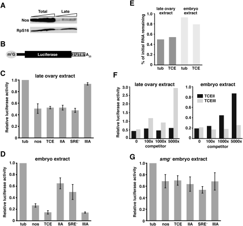
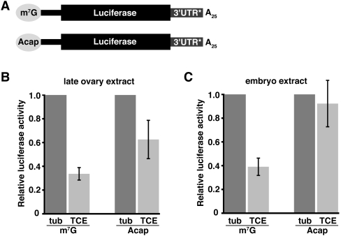
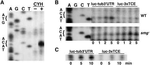
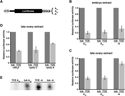
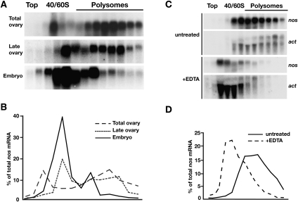
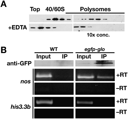
Translational control of gene expression is essential for development in organisms that rely on maternal mRNAs. In Drosophila, translation of maternal nanos (nos) mRNA must be restricted to the posterior of the early embryo for proper patterning of the anterior-posterior axis. Spatial control of nos translation is coordinated through the localization of a small subset of nos mRNA to the posterior pole late in oogenesis, activation of this localized mRNA, and repression of the remaining unlocalized nos mRNA throughout the bulk cytoplasm. Translational repression is mediated by the interaction of a cis-acting element in the nos 3' untranslated region with two proteins, Glorund (Glo) and Smaug (Smg), that function in the oocyte and embryo, respectively. The mechanism of Glo-dependent repression is unknown. Previous work suggests that Smg represses translation initiation but this model is not easily reconciled with evidence for polysome association of repressed nos mRNA. Using an in vitro translation system, we have decoupled translational repression of nos imposed during oogenesis from repression during embryogenesis. Our results suggest that both Glo and Smg regulate translation initiation, but by different mechanisms. Furthermore, we show that, during late oogenesis, nos translation is also repressed post-initiation and provide evidence that Glo mediates this event. This post-initiation block is maintained into embryogenesis during the transition to Smg-dependent regulation. We propose that the use of multiple modes of repression ensures inactivation of nos RNA that is translated at earlier stages of oogenesis and maintenance of this inactivate state throughout late oogenesis into embryogenesis.


Figures

FIGURE 1.
FIGURE 1.
In vitro translation assay for TCE-mediated repression in late ovary and embryo extracts. (A) Immunoblot of total ovary and late ovary extracts. Lanes contain increasing amounts of each extract, in twofold increments. Nos and the RpS16 loading control were detected simultaneously. (B) Architecture of the reporter RNAs used in C-G, with the m7GpppG cap, nos 5′ UTR (thinner black bar), firefly luciferase coding region, and 25-nt poly(A) tail in common. 3′ UTR* denotes sequences tested in C, D, and G: α-tubulin 3′ UTR (tub), nos 3′ UTR (nos), 3xTCE (TCE), 3xTCEIIA (IIA), 3xTCE[SRE] (SRE), or 3xTCEIIIA (IIIA). (C,D) Luciferase assays. The indicated reporter RNAs were translated in late ovary extract (C) or embryo extract (D) together with an internal control Renilla luciferase RNA. For each reaction, firefly luciferase activity of the reporter was normalized to Renilla luciferase activity. Relative luciferase activity was calculated by dividing the normalized firefly luciferase value for the indicated reporter by the normalized value for the luc-tub3′UTR reporter RNA (tub = 1.0). The graph reports the mean and standard deviation from at least three independent experiments for each reporter. (E) Quantitation of luc-tub3′UTR and luc-3xTCE RNA stability. RNA purified from in vitro translation reactions after 5 min and either 120 min (ovary) or 90 min (embryo) of incubation was analyzed by Northern blotting. The ratio of the final level to the initial level for each reporter, as determined by quantitation of the Northern blots, is plotted. (F) Competition experiment. luc-tub3′UTR and luc-3xTCE RNAs were translated in either ovary or embryo extract in the presence of 0-, 100-, 1000-, or 5000-fold molar excess of TCE stem–loop II or TCE stem–loop III RNA. Relative luciferase activity was determined as described in C and D. Similar results were obtained in multiple independent experiments. (G) Luciferase assays as described for C and D using smg mutant embryo extract.
FIGURE 2.
FIGURE 2.
Cap dependence of TCE-mediated repression. (A) Reporter RNAs used in B and C are similar to those in Figure 1 and differ from each other by the presence of an m7GpppG cap (m7G) or an ApppG cap (Acap). 3′UTR* denotes tub or 3xTCE (TCE) sequences. (B,C) Luciferase assays of the m7G and Acap luc-tub3′UTR (tub) and luc-3xTCE (TCE) reporter RNAs in late ovary or embryo extract. Relative luciferase activity was determined as described in Figure 1, except that the normalized luciferase activity of each luc-3xTCE RNA was divided by the normalized activity of its cognate luc-tub3′UTR RNA (for each, tub = 1.0).
FIGURE 3.
FIGURE 3.
The TCE decreases AUG occupancy. (A) Toeprinting experiment to monitor AUG occupancy on luc-tub3′UTR RNA in the presence (+) or absence (–) of cycloheximide (CYH). Dideoxysequencing of the reporter DNA template (four lanes on left using lighter exposures) shows the position of the initiation codon. The toeprint occurs 17 nt downstream from the A of the AUG as expected (Sachs et al. 2002). (B,C) Time-course experiments monitoring AUG toeprints on luc-tub3′UTR and luc-3xTCE reporter RNAs. (B) Reporter RNAs were translated in wild-type (WT) or smg mutant (smg) embryo extract and aliquots were removed at the indicated time points after addition of cycloheximide (at t = 0 min) for reverse transcription. (C) Toeprinting experiment performed using late ovary extract.
FIGURE 4.
FIGURE 4.
Poly(A) tail dependence of TCE-mediated repression during oogenesis. (A) Reporter RNAs used for the experiments in B–E with differences in poly(A) tail length as shown. 3′UTR* denotes tub or 3xTCE (TCE) sequences. (B–D) Luciferase assays, with relative luciferase activity determined as described for Figure 2. (B,C) Reporter RNAs shown in A were translated in late ovary or embryo extract. (D) Translation reactions containing luc-tub3′UTR (tub) and luc3xTCE (TCE) RNAs with standard 25A tails were challenged by addition of a 1000-fold molar excess of poly(C) or poly(A), or an equivalent volume of dH2O. (E) Toeprinting showing AUG occupancy for the indicated reporter RNAs in late ovary extract.
FIGURE 5.
FIGURE 5.
Temporal analysis of nos polysome association. (A) Total ovary, late ovary, and embryo extracts were fractionated on 20%–50% sucrose density gradients and RNA isolated from gradient fractions was analyzed by Northern blotting with probes for nos mRNA. (B) Quantitation of nos mRNA distribution from Northern blots in A as a % of total nos radioactivity. (C) Fractionation of late ovary extract on 20%–50% sucrose density gradients with or without addition of EDTA to disrupt polysomes. Polysomes are generally less well preserved in this experiment than in the experiment shown in A due to the lowered Mg2+ concentration in the extract necessary for polysome disruption (see Materials and Methods; Clark et al. 2000). RNA isolated from gradient fractions was analyzed by Northern blotting with probes for nos and actin mRNAs (actin migrates as two species). (D) Quantitation of nos mRNA distribution in C as a % of total nos radioactivity.
FIGURE 6.
FIGURE 6.
Glo is associated with polysomal nos mRNA in vivo. (A) Immunoblot analysis of Glo protein in fractions from the late ovary gradients shown in Figure 5C. Polysomal fractions were TCA-precipitated prior to immunoblotting, and ∼10-fold more total protein was loaded per lane. The distribution of Glo shifts to lighter fractions when polysomes are disrupted by EDTA treatment. (B) Co-immunoprecipitation of Glo and nos mRNA. A polyclonal anti-GFP antibody was used for immunoprecipitation from pelleted polysomes from wild-type (WT) or EGFP–Glo expressing late ovaries. Top panels: immunoblot analysis with a monoclonal anti-GFP antibody confirms specific purification of EGFP–Glo. Input sample is 1/10 volume equivalent of the immunoprecipitate sample (IP). Bottom panels: RT-PCR analysis of RNA isolated from immunoprecipitates detects nos but not the control his3.3b RNA. Reactions were performed with (+RT) or without (−RT) reverse transcriptase.

References

    1. Becalska AN, Gavis ER 2009. Lighting up mRNA localization in Drosophila oogenesis. Development 136: 2493–2503 - PMC - PubMed
    1. Becalska AN, Kim YR, Belletier NG, Lerit DA, Sinsimer KS, Gavis ER 2011. Aubergine is a component of a nanos mRNA localization complex. Dev Biol 349: 46–52 - PMC - PubMed
    1. Benoit P, Papin C, Kwak JE, Wickens M, Simonelig M 2008. PAP- and GLD-2-type poly(A) polymerases are required sequentially in cytoplasmic polyadenylation and oogenesis in Drosophila. Development 135: 1969–1979 - PMC - PubMed
    1. Bergsten SE, Gavis ER 1999. Role for mRNA localization in translational activation but not spatial restriction of nanos RNA. Development 126: 659–669 - PubMed
    1. Chekulaeva M, Hentze MW, Ephrussi A 2006. Bruno acts as a dual repressor of oskar translation, promoting mRNA oligomerization and formation of silencing particles. Cell 124: 521–533 - PubMed

Publication types

LinkOut - more resources