. 2011 Jul;7(7):716-26.
doi: 10.4161/auto.7.7.15155. Epub 2011 Jul 1.

The identification and analysis of phosphorylation sites on the Atg1 protein kinase

Affiliations

The identification and analysis of phosphorylation sites on the Atg1 protein kinase

Yuh-Ying Yeh et al. Autophagy. 2011 Jul.

Abstract

Autophagy is a conserved, degradative process that has been implicated in a number of human diseases and is a potential target for therapeutic intervention. It is therefore important that we develop a thorough understanding of the mechanisms regulating this trafficking pathway. The Atg1 protein kinase is a key element of this control as a number of signaling pathways target this enzyme and its associated protein partners. These studies have established that Atg1 activities are controlled, at least in part, by protein phosphorylation. To further this understanding, we used a combined mass spectrometry and molecular biology approach to identify and characterize additional sites of phosphorylation in the Saccharomyces cerevisiae Atg1. Fifteen candidate sites of phosphorylation were identified, including nine that had not been noted previously. Interestingly, our data suggest that the phosphorylation at one of these sites, Ser-34, is inhibitory for both Atg1 kinase activity and autophagy. This site is located within a glycine-rich loop that is highly conserved in protein kinases. Phosphorylation at this position in several cyclin-dependent kinases has also been shown to result in diminished enzymatic activity. In addition, these studies identified Ser-390 as the site of autophosphorylation responsible for the anomalous migration exhibited by Atg1 on SDS-polyacrylamide gels. Finally, a mutational analysis suggested that a number of the sites identified here are important for full autophagy activity in vivo. In all, these studies identified a number of potential sites of regulation within Atg1 and will serve as a framework for future work with this enzyme.


Figures

Figure 1
Figure 1
A phosphorylation site map for the S. cerevisiae Atg1. (A) The phosphorylation sites in Atg1 that were identified in this study. The 15 sites identified herein are indicated above the Atg1 schematic. All were identified by the MS/MS analysis except for S390 (indicated by a double asterisk). S515, indicated by a single asterisk, was identified previously as a site of PKA phosphorylation. The two positions indicated below the Atg1 schematic were identified previously: S508 was also found to be a site of PKA phosphorylation and T226 is a conserved site of autophosphorylation within the Atg1 kinase domain., The conserved kinase and C-terminal domains of Atg1 are indicated with the light gray (KINASE) and dark gray (CTD) shading, respectively. (B) The list of tryptic phosphopeptides identified by the tandem MS/MS analysis. The data shown include the first and last positions of the peptide, the number of phosphates present (#P), the amino acid that was phosphorylated (Mod. AA), the Mascot score, the mass deviation in ppm (Mass dev) and the charge state of each peptide (m/z). The identified sites are grouped into two categories based on the relative confidence in their respective identifications; the lower scoring sites are listed in the bottom portion of the table. Phosphorylation sites assignments, including those for the three low scoring phosphopeptides (Thr-129, Ser-533 and Ser-680), were in general supported by the observed MS/MS ions, as manually interpreted and annotated in Figure S1.
Figure 2
Figure 2
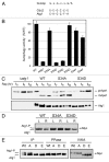
Phosphorylation at Ser-34 within Atg1 is a potential site of inhibition for the autophagy process. (A) A sequence alignment of the glycine-rich loop (G-loop) present in Atg1 and the mammalian Cdc2 enzymes. The consensus sequence for this motif is shown at the top and the residues known to be phosphorylated are underlined. (B) Assessing autophagy with the Pho8Δ60 alkaline phosphatase-based assay. The relative levels of autophagy for an atg1Δ strain carrying single-copy plasmids expressing the indicated variants of Atg1 are shown. Autophagy was induced by treating log phase cultures with 200 ng/ml rapamycin for 4 h. The data shown are the average of at least three independent experiments and the error bars indicate one standard deviation. (C) Autophagy activity as measured with the Ape1 processing assay. Cells overexpressing Ape1 were treated with 200 ng/ml rapamycin for the time indicated to induce the autophagy process. The relative level of the mature, processed form of the Ape1 protein (mApe1) was an indicator of the autophagy activity present. The bottom part shows the relative levels of the different myc-tagged Atg1 proteins in the strains examined. The electrophoretic conditions used for this latter control would not have resolved the two forms of Atg1 that comprise the doublet shown elsewhere. (D) An analysis of Atg1 autophosphorylation activity in vivo. The relative level of the autophosphorylated form of Atg1 (Atg1-P) was assessed in atg1Δ cells expressing the indicated variants of Atg1. Western blots were performed with extracts prepared from either log phase (L) or rapamycin-treated (R) cells. (E) An examination of the effects of altering Ser-34 on Atg1 autophosphorylation in vitro. The indicated myc epitope-tagged Atg1 proteins were immunoprecipitated and then either mock treated (IP) or incubated with λ phosphatase (PPase). An aliquot of the phosphatase-treated protein was then subjected to an in vitro autophosphorylation reaction (IVKA) with 2.5 mM ATP, as described in the Materials and Methods. The resulting reaction products were separated by SDS-polyacrylamide gel electrophoresis and the relative level of the autophosphorylated form of Atg1, Atg1-P, was assessed by western blotting. The (A, D and E) designations indicate the Atg1S34A, Atg1S34D and Atg1S34E proteins, respectively.
Figure 3
Figure 3
An analysis of the autophagy and kinase activities associated with alterations of the Atg1 phosphorylation sites identified in this study. (A) Autophagy activity as measured with the Pho8Δ60 alkaline phosphatase-based assay. The relative levels of autophagy present in an atg1Δ strain expressing the indicated Atg1 protein variants are shown. Autophagy was induced by treating log phase cultures with 200 ng/ml rapamycin for 4 h. The values are shown as a percentage of the activity observed with the wild-type Atg1. The data shown are the average of at least three independent experiments and the error bars indicate one standard deviation. (B) An assessment of Atg1 autophosphorylation levels in vivo. The relative levels of the autophosphorylated form (Atg1-P) of each of the indicated Atg1 variants was assessed by western blotting. The extracts were prepared from either log phase (L) or rapamycin-treated (R) cells. (C) Atg1 autophosphorylation in vitro. The indicated Atg1 proteins were immunoprecipitated from rapamycin-treated cells and subjected to an in vitro autophosphorylation assay with [γ-32P] ATP, as described in the Materials and Methods. The reaction products were separated on an SDS-polyacrylamide gel and the relative amount of radioactivity incorporated into Atg1 was assessed with a phosphorimager. The data presented represent the average of at least two independent experiments where the phosphorylation levels varied by less than 15% between the different experimental replicates.
Figure 4
Figure 4
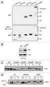
Autophosphorylation at Ser-390 is responsible for the slower-migrating form of Atg1 observed on SDS-polyacrylamide gels. (A) An in vitro autophosphorylation assay with N-terminal fragments of Atg1. The indicated Atg1 deletion constructs were immunoprecipitated from yeast cell extracts, incubated with [γ-32P] ATP and then separated on SDS-polyacrylamide gels. The relative amount of radioactivity incorporated into each Atg1 fragment was assessed by autoradiography. The lower part shows a western blot depicting the relative amount of each construct present in the kinase reactions. The positions of the relevant protein bands are indicated with an asterisk. (B) The identification of a second site of autophosphorylation within the N-terminal 420 amino acids of Atg1. Atg11-420 fragments with either a threonine (Wt) or glutamic acid (T226E) at position 226 were precipitated from cell extracts and then subjected to an in vitro autophosphorylation assay with [γ-32P] ATP. The relative amount of radioactivity incorporated into each fragment was determined by autoradiography. The lower part shows a western blot depicting the relative amount of each construct present in the kinase reactions. (C) Alterations of Ser-390 influence Atg1 mobility in an SDS-polyacrylamide gel. Western blots were performed with extracts prepared from either log phase (L) or rapamycin-treated (R) cells expressing the indicated variants of Atg1. D211A, a kinase-defective Atg1. (D) The gel mobility of the Atg1S390A and Atg1S390D variants were not affected by either a phosphatase treatment or autophosphorylation reaction. The indicated Atg1 proteins were precipitated and then either mock treated (IP) or incubated with λ phosphatase (PPase). An aliquot of the phosphatase-treated protein was then subjected to an in vitro autophosphorylation reaction (IVKA) with 2.5 mM ATP, as described in the Materials and Methods. The resulting reaction products were separated by SDS-polyacrylamide gel electrophoresis and the relative mobility on SDS-polyacrylamide gels was assessed by western blotting. The (A and D) designations indicate the Atg1S390A and Atg1S390D proteins, respectively.
Figure 5
Figure 5
Protein kinase and autophagy activities associated with alterations of Ser-390 in Atg1. (A) Atg1 autophosphorylation in vitro. The indicated Atg1 proteins were immunoprecipitated from rapamycin-treated cells and subjected to an in vitro autophosphorylation assay with [γ-32P] ATP, as described in the Materials and Methods. The reaction products were separated on an SDS-polyacrylamide gel and the relative amount of radioactivity incorporated into Atg1 was assessed with a phosphorimager. The data presented represent the average of at least two independent experiments where the phosphorylation levels varied by less than 10% between the different experimental replicates. (B) Assessing the levels of Thr-226 phosphorylation in vivo. The indicated Atg1 proteins were immunoprecipitated from rapamycin-treated cells, separated on SDS-polyacrylamide gels, and then analyzed by western blotting with the indicated antibodies. The protein signals were detected with a LI-COR Odyssey infrared detection system. The relative signal detected with the anti-pT226 phosphospecific antibody (α-pT226) is a measure of the level of autophosphorylation at Thr-226. The (A, D and E) designations indicate the Atg1S390A, Atg1S390D and Atg1S390E proteins, respectively. (C–E) Autophagy activity associated with atg1Δ cells expressing the indicated Atg1 proteins was assessed with the Pho8Δ60 processing (in C), Ape1 maturation (in D) and GFP-Atg8 processing (in E) assays, as described in the Materials and Methods. Autophagy was induced by treating log phase cells with 200 ng/ml rapamycin for 4 h (in C) or the times indicated (in D and E).

References

    1. Nakatogawa H, Suzuki K, Kamada Y, Ohsumi Y. Dynamics and diversity in autophagy mechanisms: lessons from yeast. Nat Rev Mol Cell Biol. 2009;10:458–467. - PubMed
    1. Klionsky DJ. Autophagy: from phenomenology to molecular understanding in less than a decade. Nat Rev Mol Cell Biol. 2007;8:931–937. - PubMed
    1. Todde V, Veenhuis M, van der Klei IJ. Autophagy: principles and significance in health and disease. Biochim Biophys Acta. 2009;1792:3–13. - PubMed
    1. Xie Z, Klionsky DJ. Autophagosome formation: core machinery and adaptations. Nat Cell Biol. 2007;9:1102–1109. - PubMed
    1. Yamamoto A, Cremona ML, Rothman JE. Autophagy-mediated clearance of huntingtin aggregates triggered by the insulin-signaling pathway. J Cell Biol. 2006;172:719–731. - PMC - PubMed

Publication types

MeSH terms