Comparative Study
. 2011 May;12(5):463-9.
doi: 10.1038/embor.2011.43. Epub 2011 Apr 1.

The oncometabolite 2-hydroxyglutarate inhibits histone lysine demethylases

Affiliations
Comparative Study

The oncometabolite 2-hydroxyglutarate inhibits histone lysine demethylases

Rasheduzzaman Chowdhury et al. EMBO Rep. 2011 May.

Abstract

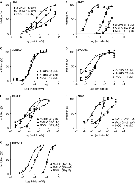
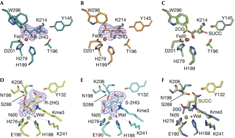
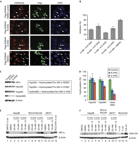
Mutations in isocitrate dehydrogenases (IDHs) have a gain-of-function effect leading to R(-)-2-hydroxyglutarate (R-2HG) accumulation. By using biochemical, structural and cellular assays, we show that either or both R- and S-2HG inhibit 2-oxoglutarate (2OG)-dependent oxygenases with varying potencies. Half-maximal inhibitory concentration (IC(50)) values for the R-form of 2HG varied from approximately 25 μM for the histone N(ɛ)-lysine demethylase JMJD2A to more than 5 mM for the hypoxia-inducible factor (HIF) prolyl hydroxylase. The results indicate that candidate oncogenic pathways in IDH-associated malignancy should include those that are regulated by other 2OG oxygenases than HIF hydroxylases, in particular those involving the regulation of histone methylation.


Conflict of interest statement

The authors declare that they have no conflict of interest.

Figures

Figure 1
Figure 1
Inhibition of recombinant forms of 2OG oxygenases by R-/S-2HG and N-oxalylglycine. (A) Inhibition of FIH as assessed by measuring hydroxylation of HIF1αCAD using a MALDI–TOF–MS assay. (B) Inhibition of PHD2 as assessed by measuring hydroxylation of HIF1αCODD using a MALDI–TOF–MS assay. Inhibition of (C) JMJD2A, (D) JMJD2C, (E) FBXL11 and (F) ABH2 by R-, S-2HG and NOG, as assessed by using formaldehyde dehydrogenase-based assay. (G) Inhibition of BBOX1 by S-2HG and R-2HG as compared with the standard NOG by NMR. All assays were performed in triplicate and are shown as averages with s.e.m. IC50 values were calculated using log(inhibitor) compared with normalized response (variable slope), using the program Prism, and are given in parentheses. The assay conditions for each enzyme are described in supplementary Table S1 online. ABH2, AlkB homologue 2; BBOX1, γ-butyrobetaine hydroxylase 1; FIH, factor inhibiting-hypoxia-inducible factor; HIF, hypoxia-inducible factor; 2OG, 2-oxoglutarate; PHD2, prolyl hydroxylase 2; R-2HG, R-enantiomer of 2-hydroxyglutarate; IC50, half-maximal inhibitory concentration; MALDI–TOF–MS, matrix-assisted laser desorption/ionization–time of flight–mass spectrometry; NOG, N-oxalylglycine.
Figure 2
Figure 2
Crystal structures of FIH and JMJD2A in complex with R- and S-2HG indicate binding of 2HG to the 2OG-binding site. FIH/JMJD2A folds were similar to those reported (RMSD for Cα⩽0.3 Å for monomers). Dotted lines indicate apparent hydrogen bonds and polar interactions. Data collection and refinement statistics are given in supplementary Table S2 online. (A) View of the FIH.Fe(II).R-2HG complex showing the Fo–Fc omit map (contoured to 6 σ) for R-2HG. Notably, the side chain of Trp 296FIH, which is involved in substrate binding (Elkins et al, 2003), has two conformations. (B) FIH.Fe(II).S-2HG structure showing the Fo–Fc omit map contoured to 6 σ around S-2HG. (C) Overlay of views from structures of FIH in complex with 2OG or succinate. (D) View from the active site of the JMJD2A.Ni(II).R-2HG.H3K36me3 structure showing the Fo–Fc omit map contoured to 3.5 σ around R-2HG; Kme3=Nε-trimethylated lysine substrate residue; Ni(II) substitute for Fe(II). (E) JMJD2A.Ni(II).S-2HG.H3K36me3 structure showing the Fo–Fc omit map contoured to 3.5 σ around S-2HG. (F) Overlay of views from structures of JMJD2A in complex with 2OG or succinate. (DF) Both R-/S-2HG induce a rotation of approximately 100° (relative to other JMJD2A structures) about Cα-Cβ of the Ser 288JMJD2A side chain such that it is positioned to hydrogen bond to 2HG (Ser Oγ-R/S-2HG O2; 2.8/3.1 Å). Ser 288JMJD2A is proposed to be involved in substrate recognition and a determinant of the specificity of JMJD2 for different methylation states (Chen et al, 2006). FIH, factor inhibiting-hypoxia-inducible factor; 2OG, 2-oxoglutarate; R-2HG, R-enantiomer of 2-hydroxyglutarate; RMSD, root mean square deviation.
Figure 3
Figure 3
Inhibition of histone demethylases and HIF hydroxylases by R- and S-2HG. (A,B) Inhibition of JMJD2A activity in HeLa cells, as shown by immunofluorescence studies. JMJD2A was expressed as an N-terminally Flag-tagged protein. The results for DMOG, a cell-penetrating form of the ‘generic’ 2OG oxygenase inhibitor NOG, are shown for comparison. (A) Indirect immunofluorescence analyses using antibodies against H3K9me3 (left panels) or Flag (middle panels) were used to analyse the inhibition of ectopically expressed JMJD2A. DAPI staining (right panels) indicates location of nuclei. Rows represent control (0.5% DMSO), R-/S-2HG- and DMOG-treated cells. White arrows in the left and right panels indicate nuclei in which JMJD2A is overexpressed. Higher levels of H3K9me3 were detected in both R- and S-2HG-treated cells, indicating inhibition of JMJD2A, whereas the control shows only a level of H3K9me3 in nuclei in which JMJD2A is overexpressed. (B) Percentage inhibition of JMJD2A activity, using addition of doses of R- and S-2HG in their dimethyl ester forms (5 or 10 mM × 8); error bars represent standard error (n>50). (C,D) Effect of R-/S-2HG on HIF1α hydroxylation in VHL-defective RCC4/VA cells. (C) Immunoblots of protein extracts from RCC4 cells treated for 4 h with R- and S-2HG compared with a DMSO-treated control. The compounds were added using the same dosing regime (10 mM × 8) as was used for the JMJD2A cell-work in their dimethyl ester forms. Signals of total HIF1α and hydroxylated NODD, CODD and CAD were analysed. (D) Quantification of data from (C) by densitometry; band intensities were normalized to HIF1α signals. Vertical bars represent standard errors (n>3). (E) Effect of R-/S-2HG on HIF1α protein levels in human hepatoma (Hep3B), renal (RCC4/VHLHA, overexpressing haemagglutinin-tagged VHL) and breast cancer (MCF7) cells. (F) Immunoblot analyses of HIF1α CAD hydroxylation of selected samples from (E). DMOG, dimethyloxalylglycine; HIF, hypoxia-inducible factor; NOG, N-oxalylglycine; RCC, renal cell carcinoma; R-2HG, R-enantiomer of 2-hydroxyglutarate; 2OG, 2-oxoglutarate.

References

    1. Chen Z et al. (2006) Structural insights into histone demethylation by JMJD2 family members. Cell 125: 691–702 - PubMed
    1. Chowdhury R, McDonough MA, Mecinovic J, Loenarz C, Flashman E, Hewitson KS, Domene C, Schofield CJ (2009) Structural basis for binding of hypoxia-inducible factor to the oxygen-sensing prolyl hydroxylases. Structure 17: 981–989 - PubMed
    1. Conejo-Garcia A, McDonough MA, Loenarz C, McNeill LA, Hewitson KS, Ge W, Lienard BM, Schofield CJ, Clifton IJ (2010) Structural basis for binding of cyclic 2-oxoglutarate analogues to factor-inhibiting hypoxia-inducible factor. Bioorg Med Chem Lett 20: 6125–6128 - PubMed
    1. Couture JF, Collazo E, Ortiz-Tello PA, Brunzelle JS, Trievel RC (2007) Specificity and mechanism of JMJD2A, a trimethyllysine-specific histone demethylase. Nat Struct Mol Biol 14: 689–695 - PubMed
    1. Dalgliesh GL et al. (2010) Systematic sequencing of renal carcinoma reveals inactivation of histone modifying genes. Nature 463: 360–363 - PMC - PubMed

Publication types

MeSH terms