. 2011 Jul;8(4):305-14.
doi: 10.1038/cmi.2011.8. Epub 2011 Apr 4.

The increase in surface CXCR4 expression on lung extravascular neutrophils and its effects on neutrophils during endotoxin-induced lung injury

Affiliations

The increase in surface CXCR4 expression on lung extravascular neutrophils and its effects on neutrophils during endotoxin-induced lung injury

Mitsuhiro Yamada et al. Cell Mol Immunol. 2011 Jul.

Abstract

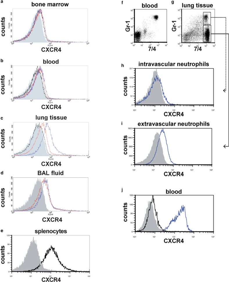
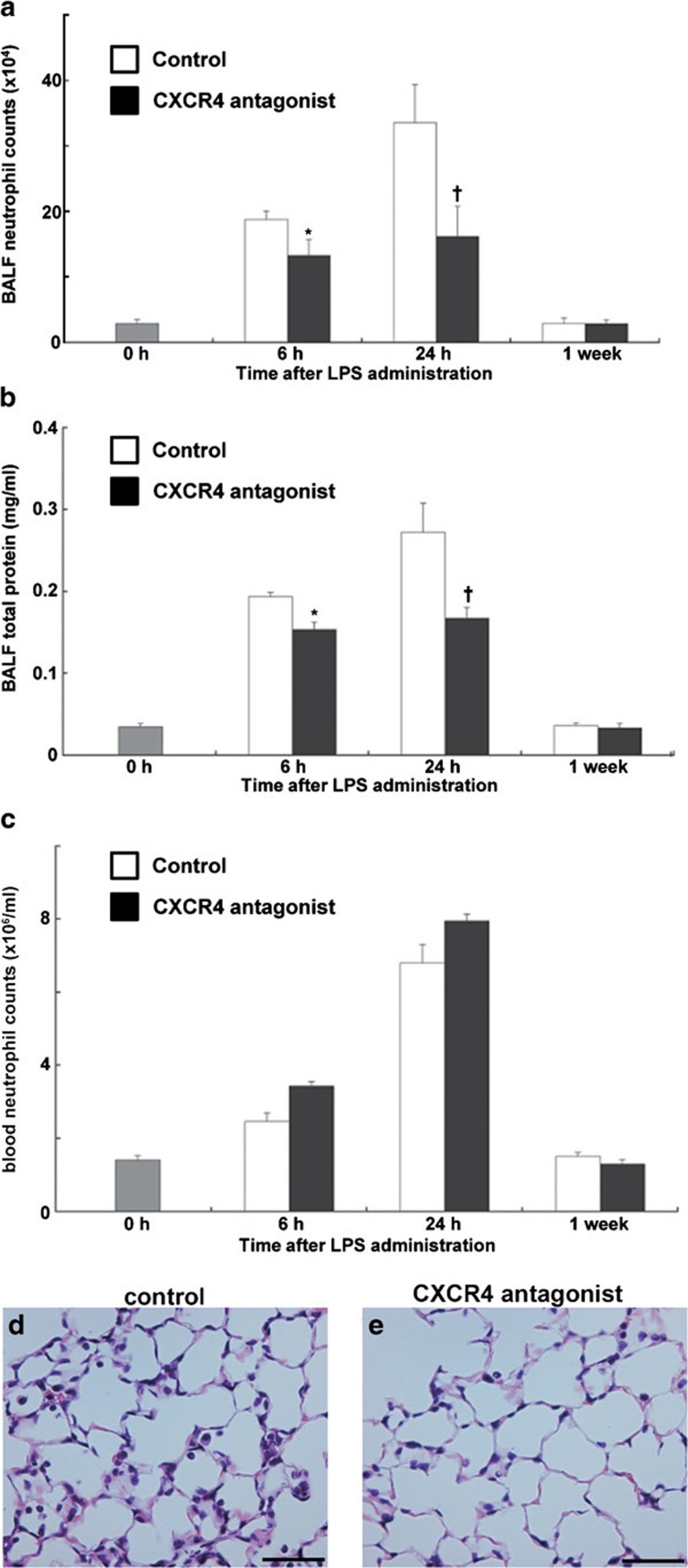
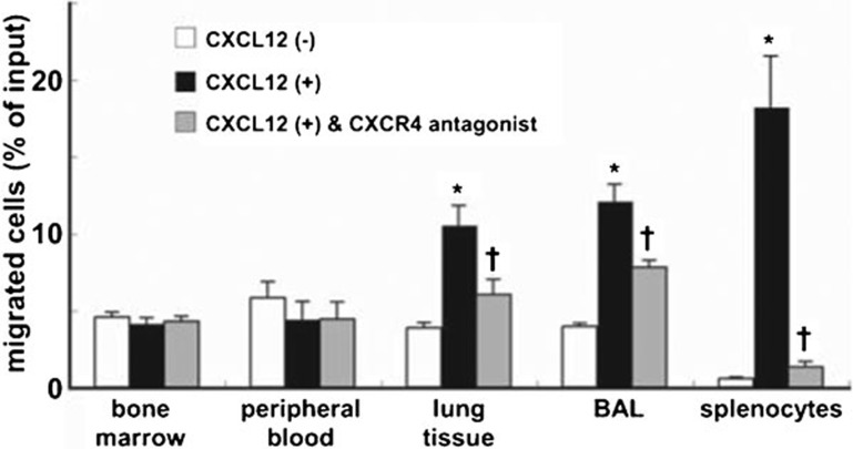
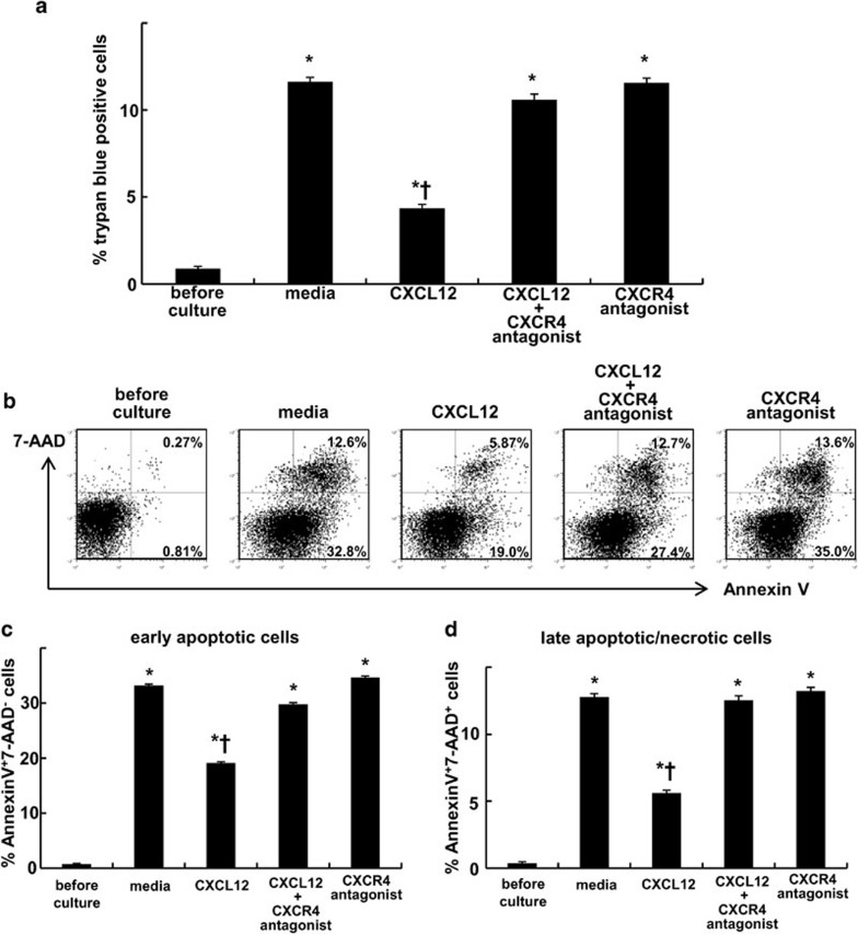
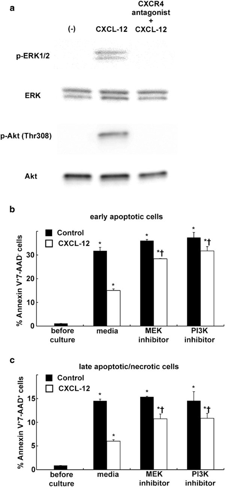
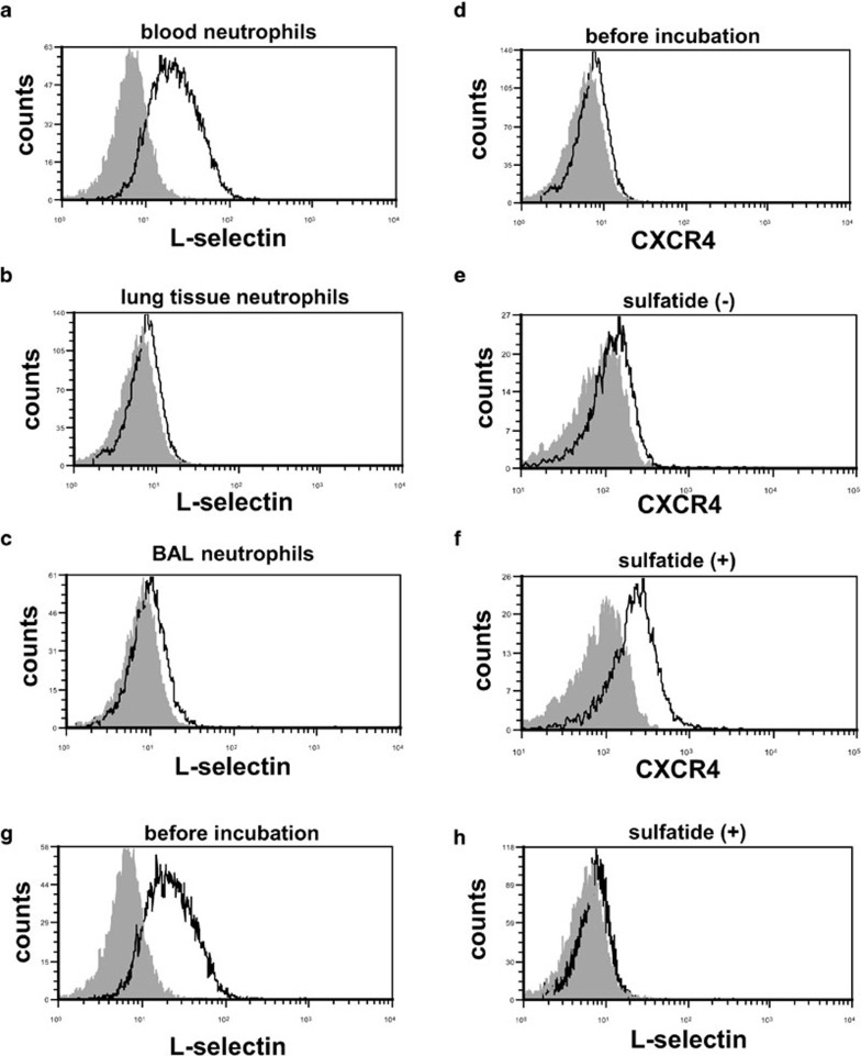
Inflammatory stimuli, such as a microbes or lipopolysaccharides, induce a rapid release of neutrophils from the bone marrow and promote neutrophil migration into inflamed sites to promote host defense. However, an excess accumulation and retention of neutrophils in inflamed tissue can cause severe tissue injuries in the later stages of inflammation. Recent studies have reported that both CXCL12 levels in injured lungs and its receptor, CXCR4, on accumulated neutrophils in injured lungs, increased; furthermore, these studies showed that the CXCL12/CXCR4 signaling pathway participated in neutrophil accumulation in the later stages of lipopolysaccharide (LPS)-induced lung injury. However, the mechanisms underlying this increase in surface CXCR4 expression in neutrophils remain unclear. In this study, we found that surface CXCR4 expression increased in extravascular, but not intravascular, neutrophils in the lungs of LPS-induced lung injury model mice. Furthermore, ex vivo studies revealed that CXCL12 acted not only as a chemoattractant, but also as a suppressor of cell death for the lung neutrophils expressing CXCR4. Sulfatide, one of the native ligands for L-selectin, induced the increase of surface CXCR4 expression on isolated circulating neutrophils, suggesting that the activation of L-selectin may be involved in the increase in surface CXCR4. Our findings show that surface CXCR4 levels on neutrophils increase after extravasation into injured lungs, possibly through the activation of L-selectin. The CXCL12/CXCR4 signaling pathway plays an important role in the modulation of neutrophil activity during acute lung injury, not only by promoting chemotaxis but also by suppressing cell death.


Figures

Figure 1
Figure 1
Surface CXCR4 expression increased in extravascular neutrophils in the mouse lungs during LPS-induced lung injury. (ae) Flow cytometric analyses were performed to determine the surface CXCR4 expression levels of neutrophils isolated from bone marrow (a), peripheral blood (b), lung tissue (c) and BAL fluid (d). Neutrophils were analyzed before LPS administration (black line) and at 6 h (red line) and 24 h (blue line) afterward. The filled images show the staining using an isotype-matched control antibody. We were unable to analyze the surface CXCR4 expression levels of neutrophils isolated from BAL fluid before LPS administration because there were an insufficient number of neutrophils in these samples. Splenocytes from untreated control mice were used as a positive control for CXCR4 staining (e). (fi) The surface CXCR4 expression levels of intravascular neutrophils (g; GR-1+7/4+) and extravascular neutrophils (g; GR-17/4+) in the lungs at 24 h during LPS-induced lung injury were examined. Note almost all circulating neutrophils (7/4+ cells) in blood were stained with Gr-1 5 min after antibody injection (f). The blue lines show the surface CXCR4 expression levels of intravascular neutrophils (h) or extravascular neutrophils (i). The filled images show the staining using an isotype-matched control antibody. (h) The levels of intracellular CXCR4 (blue line) in neutrophils isolated from mouse blood were analyzed by staining with permeabilization. The filled image shows the staining using an isotype-matched control antibody. The black line shows the surface CXCR4 expression levels. Representative histograms or dot plots from one of three experiments that showed similar results are presented. BAL, bronchoalveolar lavage; LPS, lipopolysaccharide.
Figure 2
Figure 2
Blocking the CXCL12/CXCR4 signaling pathway inhibited neutrophil migration into the lung air space and the increase of lung permeability during LPS-induced lung injury. (ac) Neutrophil counts (a), total protein concentration (b) in the BALF and neutrophil counts in the circulating blood (c) were determined in C57BL/6 mice treated with either PBS (white) or a CXCR4 antagonist (black) at indicated time points during LPS-induced lung injury. A total of six mice were used in each group. Values represent mean±s.e.m. *P<0.01, P<0.05, versus PBS control mice using ANOVA with Scheffé's post hoc test. (d, e) Histological evaluation of the treatment with a CXCR4 antagonist on LPS-induced lung injury. Representative images of hematoxylin and eosin stained lung tissue sections from PBS- (d) or CXCR4 antagonist-treated (e) mice at 24 h during LPS-induced lung injury. Scale bar=50 µm. BALF, bronchoalveolar lavage fluid; LPS, lipopolysaccharide; PBS, phosphate-buffered saline.
Figure 3
Figure 3
Neutrophils that accumulated in the mouse lungs during LPS-induced lung injury showed migratory responses to CXCL12. Neutrophils were isolated from the bone marrow, blood, lung tissue and BAL fluid at 24 h during LPS-induced lung injury. Cell migration assays assessing the migration of neutrophils toward CXCL12 were performed in vitro using chemotaxis chambers. Splenocytes from untreated control mice were used as a positive control because splenocytes expressed high levels of surface CXCR4 (Figure 1e). Levels of neutrophil or splenocyte migration in the absence of CXCL12, the presence of CXCL12, or after pretreatment with a CXCR4 antagonist in the presence of CXCL12, are depicted by the white, black and gray bars, respectively. The data shown represent the percentage of migration. The results were obtained from five mice in each group. Values represent mean±s.e.m. *P<0.01 versus CXCL12 (−) control group and P<0.01 versus CXCL12 (+) group using ANOVA with Scheffé's post hoc test. BAL, bronchoalveolar lavage.
Figure 4
Figure 4
CXCR4 activation by CXCL12 attenuated the cell death of mouse neutrophils isolated from the injured lungs. Neutrophils were isolated from the mouse lungs at 24 h after LPS instillation. The isolated neutrophils were cultured in either media alone (RPMI 1640 supplemented with 10% FCS), media containing CXCL12, media containing CXCL12 and a specific CXCR4 antagonist or media with a specific CXCR4 antagonist alone for 24 h at 37 °C. Subsequently, cell death was assessed by trypan blue staining (a). Staining with Annexin V and 7-AAD was also performed to detect early apoptotic (Annexin V+7-AAD; b, c) cells and late apoptotic/necrotic (Annexin V+7-AAD+; b, d) cells. Representative dot plots are shown. A total of six mice were used in each group. The values represent mean±s.e.m. *P<0.01 versus before culture group and P<0.01 versus media only group using ANOVA with Scheffé's post hoc test. 7-AAD, 7-aminoactinomycin D; FCS, fetal calf serum.
Figure 5
Figure 5
CXCL12 protects lung-accumulated neutrophils of LPS-injured mice from apoptosis through MEK/ERK and PI3K/Akt pathways. (a) Neutrophils were isolated from the mouse lungs at 24 h after LPS instillation. Isolated neutrophils were untreated or pre-incubated with a CXCR4 antagonist then treated CXCL12. Western blot analysis showed that CXCL12 induced the phosphorylation of ERK1/2 and Akt. (b, c) The isolated neutrophils were cultured for 24 h at 37 °C in the presence (white bar) or absence (black bar) of CXCL12 alone or in combination with a MEK1/2 inhibitor (U0126) or a PI3K inhibitor (LY294002). Subsequently, staining with Annexin V and 7-AAD was also performed to detect early apoptotic (Annexin V+7-AAD; b) cells and late apoptotic/necrotic (Annexin V+7-AAD+; c) cells. Note the suppression of the inhibitory effect of CXCL12 against apoptosis in the presence of MEK1/2 inhibitor or PI3K inhibitor. The values represent mean±s.e.m. (n=6). *P<0.01 versus before culture group and P<0.01 versus media only group using ANOVA with Scheffé's post hoc test. 7-AAD, 7-aminoactinomycin D; LPS, lipopolysaccharide.
Figure 6
Figure 6
L-selectin may be involved in the increase in surface CXCR4 expression in mouse neutrophils. (ac) The surface L-selectin expression levels of neutrophils isolated from blood (a), lung tissue (b) and BAL fluid (c) at 24 h during LPS-induced lung injury were assessed by flow cytometry. The filled images show the staining using an isotype-matched control antibody. (df) Sulfatide induced the increase in surface CXCR4 expression in neutrophils isolated from mouse blood. The surface CXCR4 expression levels were examined before incubation (d) and after incubation for 1 h either with (f) or without (e) sulfatide. (g, h) Sulfatide induced shedding of L-selectin on neutrophils. Surface expressions of L-selectin on neutrophils were examined before incubation (g) and after incubation for 1 h with sulfatide (h). Representative histograms from one of three experiments that showed similar results are presented. BAL, bronchoalveolar lavage; LPS, lipopolysaccharide.

References

    1. Kubo H, Graham L, Doyle NA, Quinlan WM, Hogg JC, Doerschuk CM. Complement fragment-induced release of neutrophils from bone marrow and sequestration within pulmonary capillaries in rabbits. Blood. 1998;92:283–290. - PubMed
    1. Kubo H, Morgenstern D, Quinian WM, Ward PA, Dinauer MC, Doerschuk CM. Preservation of complement-induced lung injury in mice with deficiency of NADPH oxidase. J Clin Invest. 1996;97:2680–2684. - PMC - PubMed
    1. Strieter RM, Kunkel SL, Bone RC. Role of tumor necrosis factor-alpha in disease states and inflammation. Crit Care Med. 1993;21:S447–S463. - PubMed
    1. Gabay C, Lamacchia C, Palmer G. IL-1 pathways in inflammation and human diseases. Nat Rev Rheumatol. 2010;6:232–241. - PubMed
    1. Romagnani P, Lasagni L, Annunziato F, Serio M, Romagnani S. CXC chemokines: the regulatory link between inflammation and angiogenesis. Trends Immunol. 2004;25:201–209. - PubMed

Publication types

MeSH terms