

Ruthenium(II) Complex Enantiomers as Cellular Probes for Diastereomeric Interactions in Confocal and Fluorescence Lifetime Imaging Microscopy
- PMID: 21461029
- PMCID: PMC3065721
- DOI: 10.1021/jz101580e
Ruthenium(II) Complex Enantiomers as Cellular Probes for Diastereomeric Interactions in Confocal and Fluorescence Lifetime Imaging Microscopy
Abstract
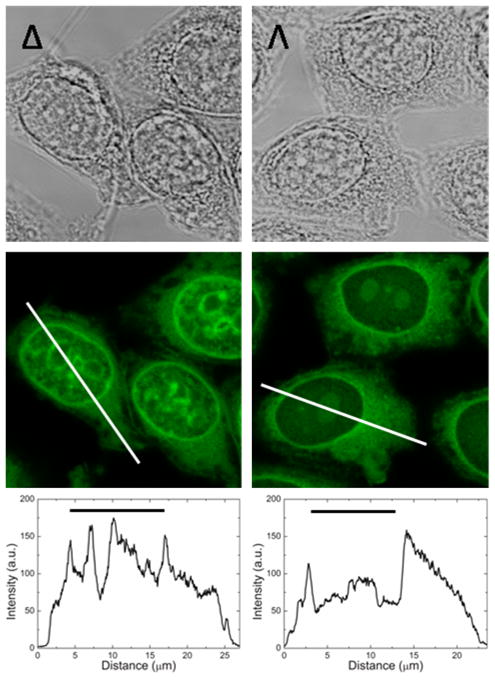
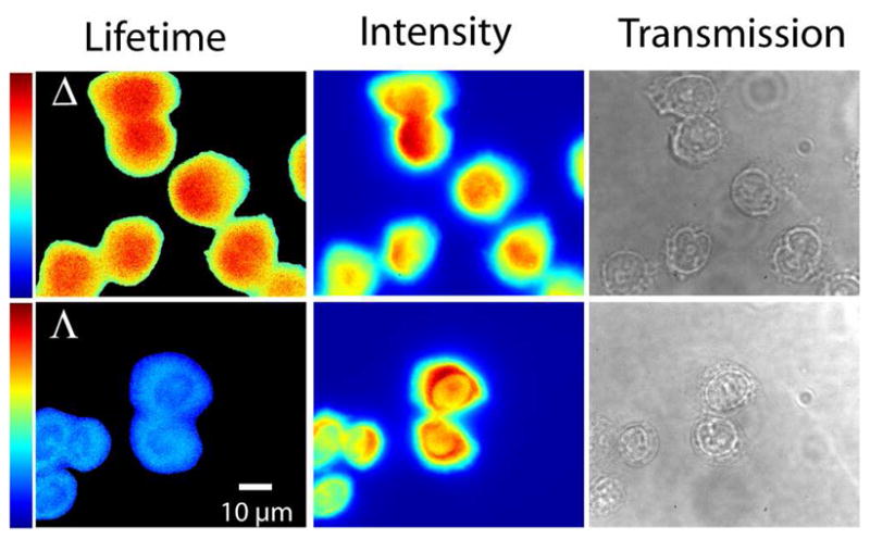
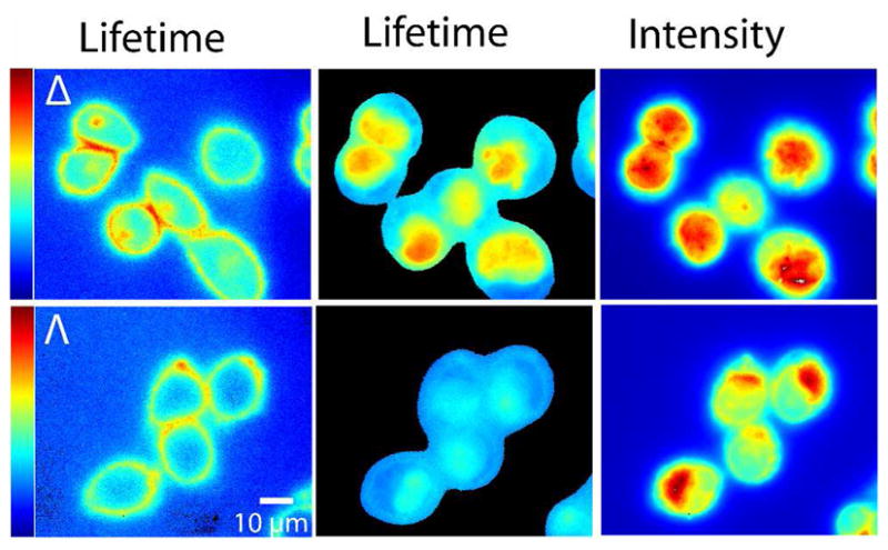
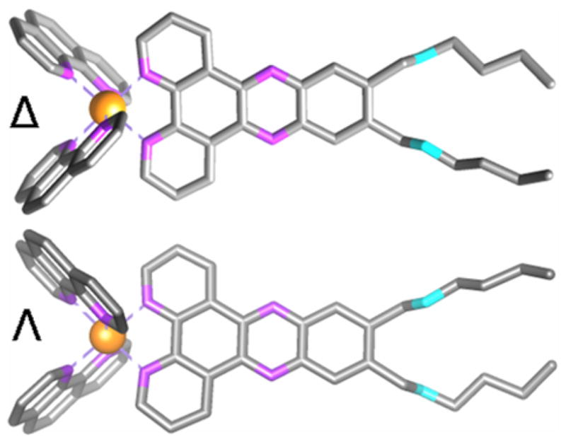
Ruthenium dipyridophenazine (dppz) complexes are sensitive luminescent probes for hydrophobic environments. Here, we apply multiple-frequency fluorescence lifetime imaging microscopy (FLIM) to Δ and Λ enantiomers of lipophilic ruthenium dppz complexes in live and fixed cells, and their different lifetime staining patterns are related to conventional intensity-based microscopy. Excited state lifetimes of the enantiomers determined from FLIM measurements correspond well with spectroscopically measured emission decay curves in pure microenvironments of DNA, phospholipid membrane or a model protein. We show that FLIM can be applied to monitor the long-lived excited states of ruthenium complex enantiomers and, combined with confocal microscopy, give new insight into their biomolecular binding and reveal differences in the microenvironment probed by the complexes.
Figures
References
-
- Barton JK, Danishefsky AT, Goldberg JM. Tris(Phenanthroline)Ruthenium(II) - Stereoselectivity in Binding to DNA. J Am Chem Soc. 1984;106:2172–2176.
-
- Brennaman MK, Meyer TJ, Papanikolas JM. [Ru(bpy)2dppz]2+ Light-Switch Mechanism in Protic Solvents as Studied Through Temperature-Dependent Lifetime Measurements. J Phys Chem A. 2004;108:9938–9944.
-
- Coates CG, Olofsson J, Coletti M, McGarvey JJ, Önfelt B, Lincoln P, Nordén B, Tuite E, Matousek P, Parker AW. Picosecond Time-Resolved Resonance Raman Probing of the Light-Switch States of [Ru(Phen)2dppz]2+ J Phys Chem B. 2001;105:12653–12664.
-
- Fernandez-Moreira V, Thorp-Greenwood FL, Coogan MP. Application of d6 Transition Metal Complexes in Fluorescence Cell Imaging. Chem Commun. 2010;46:186–202. - PubMed
-
- Jenkins Y, Friedman AE, Turro NJ, Barton JK. Characterization of Dipyridophenazine Complexes of Ruthenium(II) - the Light Switch Effect as a Function of Nucleic-Acid Sequence and Conformation. Biochemistry. 1992;31:10809–10816. - PubMed
Grants and funding
LinkOut - more resources
Full Text Sources
