Changes of Alpha1-Antitrypsin Levels in Allergen-induced Nasal Inflammation
- PMID: 21461061
- PMCID: PMC3062225
- DOI: 10.3342/ceo.2011.4.1.33
Changes of Alpha1-Antitrypsin Levels in Allergen-induced Nasal Inflammation
Abstract
Objectives: Alpha1-antitrypsin (AAT) is the main inhibitor of human neutrophil elastase, and plays a role in counteracting the tissue damage caused by elastase in local inflammatory conditions. The study evaluated the involvement of AAT in nasal allergic inflammation.
Methods: Forty subjects with mono-sensitization to Dermatophagoides pteronyssinus (Dpt) were enrolled. Twenty allergic rhinitis patients frequently complained of nasal symptoms such as rhinorrhea, stuffiness, sneezing, and showed positive responses to the nasal provocation test (NPT) with Dpt (Group I). The other 20 asymptomatic patients showed sensitization to Dpt but negative NPT (Group II). The levels of AAT, eosinophil cationic protein (ECP), and Dpt-specific IgA antibodies were measured in the nasal lavage fluids (NLFs), collected at baseline, 10 minutes, 30 minutes, 3 hours, and 6 hours after the NPT. Nasal mucosa AAT expression was evaluated with immunohistochemical staining from Group I and Group II.
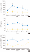
Results: At baseline, only the Dpt-specific IgA level was significantly increased in the NLFs of Group I compared with Group II, while ECP and AAT levels were not significantly different between two groups. After Dpt provocation, AAT, ECP, and Dpt-specific IgA levels were significantly increased in the NLFs of Group I during the early and late responses. The protein expression level of AAT was mostly found in the infiltrating inflammatory cells of the nasal mucosa, which was significantly increased in Group I compared to Group II.
Conclusion: The increment of AAT showed a close relationship with the activation of eosinophils induced by allergen-specific IgA in the NLFs of patients with allergic rhinitis after allergen stimulation. These findings implicate AAT in allergen-induced nasal inflammation.
Keywords: Allergic rhinitis; Alpha1-antitrypsin; Eosinophil cationic protein; IgA; Nasal lavage fluid.
Conflict of interest statement
No potential conflict of interest relevant to this article are reported.
Figures


References
-
- Naclerio RM, Proud D, Peters SP, Silber G, Kagey-Sobotka A, Adkinson NF, Jr, et al. Inflammatory mediators in nasal secretions during induced rhinitis. Clin Allergy. 1986 Mar;16(2):101–110. - PubMed
-
- Borregaard N, Cowland JB. Granules of the human neutrophilic polymorphonuclear leukocyte. Blood. 1997 May 15;89(10):3503–3521. - PubMed
-
- Travis J, Salvesen GS. Human plasma proteinase inhibitors. Annu Rev Biochem. 1983;52:655–709. - PubMed
-
- Hubbard R, Crystal RG. Antiproteases. In: Crystal RG, West JB, editors. The lung. New York: Raven Press; 1991. pp. 1775–1788.
-
- Cichy J, Potempa J, Travis J. Biosynthesis of alpha1-proteinase inhibitor by human lung-derived epithelial cells. J Biol Chem. 1997 Mar 28;272(13):8250–8255. - PubMed
LinkOut - more resources
Full Text Sources
Research Materials
Miscellaneous

