A case of autoimmune progesterone dermatitis misdiagnosed as allergic contact dermatitis
- PMID: 21461257
- PMCID: PMC3062796
- DOI: 10.4168/aair.2011.3.2.141
A case of autoimmune progesterone dermatitis misdiagnosed as allergic contact dermatitis
Abstract
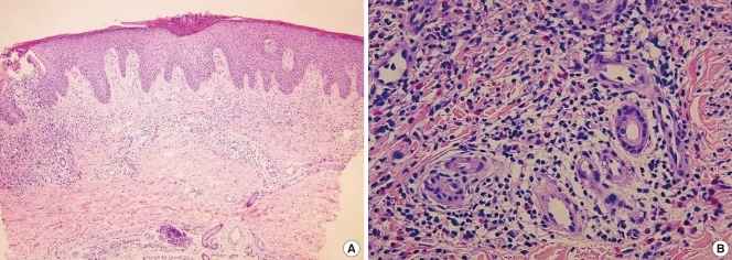
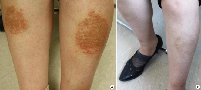
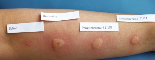
Autoimmune progesterone dermatitis is a rare autoimmune response to endogenous progesterone that usually occurs in fertile females. Cutaneous or mucosal lesions develop cyclically during the luteal phase of the menstrual cycle when progesterone levels are elevated. Symptoms usually start 3-10 days before menstruation and resolve 1-2 days after menstruation ceases. We report the case of a 48-year-old woman with intermittent eczematous skin lesions of the legs, forearms, and buttocks. She was diagnosed with allergic contact dermatitis, and topical steroids were prescribed. Her skin eruptions waxed and waned for 6 years and were associated with her menstrual cycle. We performed an intradermal test using progesterone, which was positive, and prescribed gonadotropin-releasing hormone analogues monthly for 3 months. The patient's skin lesions improved, confirming the diagnosis. Autoimmune progesterone dermatitis should be included in the differential diagnosis of recurrent eczema that is refractory to treatment in women of child-bearing age.
Keywords: Autoimmune progesterone dermatitis; allergic contact dermatitis; eczema; gonadotropin-releasing hormone analogues; intradermal test.
Conflict of interest statement
There are no financial or other issues that might lead to conflict of interest.
Figures
References
-
- Géber H. IV. Einige Daten zur Pathologie der Urticaria menstruationalis. Dermatologische Zeitschrift. 1921;32:143–150.
-
- Snyder JL, Krishnaswamy G. Autoimmune progesterone dermatitis and its manifestation as anaphylaxis: a case report and literature review. Ann Allergy Asthma Immunol. 2003;90:469–477. - PubMed
-
- Hur W, Chun SI. Autoimmune progesterone dermatitis. Korean J Dermatol. 1993;31:775–779.
-
- Lee CW, Yoon KB, Yi JU, Cho SH. Autoimmune progesterone dermatitis. J Dermatol. 1992;19:629–631. - PubMed
-
- Wilkinson SM, Beck MH, Kingston TP. Progesterone-induced urticaria--need it be autoimmune? Br J Dermatol. 1995;133:792–794. - PubMed