. 2011 Apr 1;34(4):425-34.
doi: 10.1093/sleep/34.4.425.

Phasic motor activity of respiratory and non-respiratory muscles in REM sleep

Affiliations

Phasic motor activity of respiratory and non-respiratory muscles in REM sleep

Jimmy J Fraigne et al. Sleep. .

Abstract

Objectives: In this study, we quantified the profiles of phasic activity in respiratory muscles (diaphragm, genioglossus and external intercostal) and non-respiratory muscles (neck and extensor digitorum) across REM sleep. We hypothesized that if there is a unique pontine structure that controls all REM sleep phasic events, the profiles of the phasic twitches of different muscle groups should be identical. Furthermore, we described how respiratory parameters (e.g., frequency, amplitude, and effort) vary across REM sleep to determine if phasic processes affect breathing.

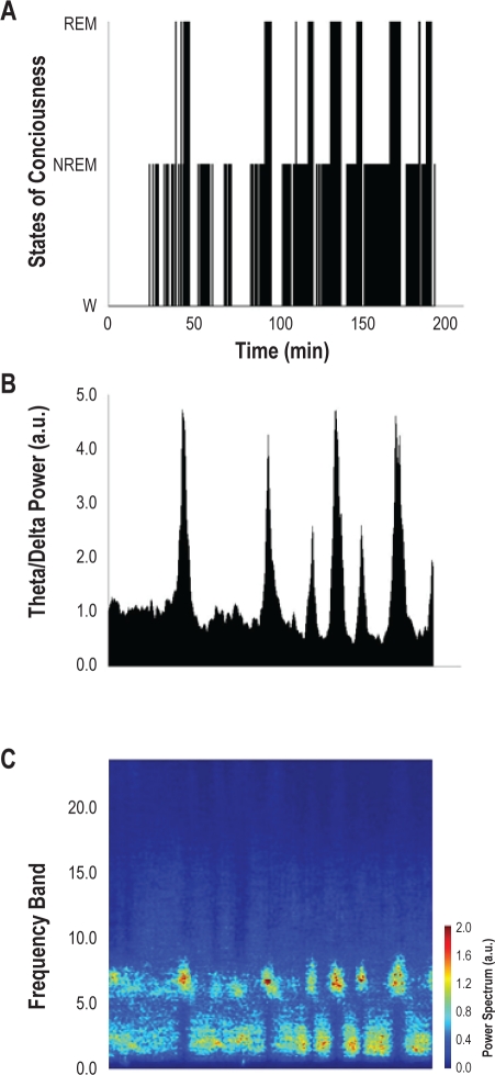
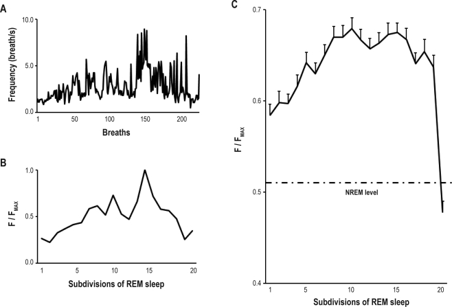
Methods: Electrodes were implanted in Wistar rats to record brain activity and muscle activity of neck, extensor digitorum, diaphragm, external intercostal, and genioglossal muscles. Ten rats were studied to obtain 313 REM periods over 73 recording days. Data were analyzed offline and REM sleep activity profiles were built for each muscle. In 6 animals, respiratory frequency, effort, amplitude, and inspiratory peak were also analyzed during 192 REM sleep periods.

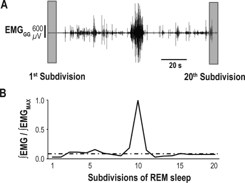
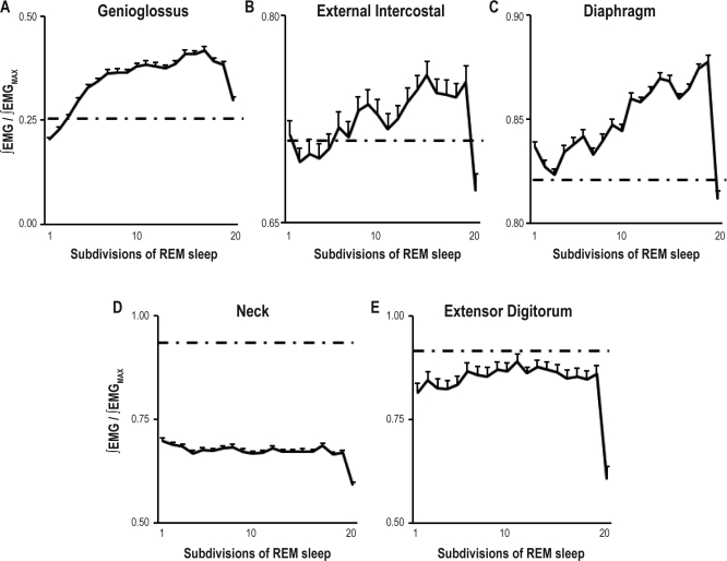
Results: Respiratory muscle phasic activity increased in the second part of the REM period. For example, genioglossal activity increased in the second part of the REM period by 63.8% compared to the average level during NREM sleep. This profile was consistent between animals and REM periods (η(2)=0.58). This increased activity seen in respiratory muscles appeared as irregular bursts and trains of activity that could affect rythmo-genesis. Indeed, the increased integrated activity seen in the second part of the REM period in the diaphragm was associated with an increase in the number (28.3%) and amplitude (30%) of breaths. Non-respiratory muscle phasic activity in REM sleep did not have a profile like the phasic activity of respiratory muscles. Time in REM sleep did not have an effect on nuchal activity (P=0.59).

Conclusion: We conclude that the concept of a common pontine center controlling all REM phasic events is not supported by our data. There is a drive in REM sleep that affects specifically respiratory muscles. The characteristic increase in respiratory frequency during REM sleep is induced by this drive.

Keywords: REM phasic event; diaphragm; external intercostal; genioglossus; rat.


Figures

Figure 1
Figure 1
Continuous recording of genioglossal muscle activity (EMGGG), nuchal muscle activity (EMGN), and EEG during NREM sleep, REM sleep, and wakefulness.
Figure 2
Figure 2
Sleep and wakefulness in a 3-h session in one animal (MST7). (A) Hypnogram; (B) Theta power to delta power ratio as a function of time for the same session as in A; (C) Power spectrum of various frequency bands (absolute values) as a function of time for the same session as in A.
Figure 3
Figure 3
Analysis of rectified EMG in 20 subdivisions. (A) Recording of genioglossal (EMGGG) muscle activity during a REM period. (B) EMGGG is rectified then integrated for each of the 20 subdivisions. The activity is expressed as a ratio of the integrated EMG over the maximum activity during the REM period. Average ∫EMG level during the preceding NREM sleep period is indicated as an intermittent line. The integrated activity across all 20 subdivisions, with repeated measurements over all REM periods, was analyzed then with an ANOVA (not shown).
Figure 4
Figure 4
Genioglossus (A), external intercostal (B), diaphragm (C), neck (D), and extensor digitorum (E) muscle activity across 313 REM periods divided into 20 subdivisions from 10 Wistar rats. Activity is expressed as a ratio of activity to the REM sleep peak activity for each muscle and each REM period. Average ∫EMG level during the preceding NREM sleep period is indicated as an intermittent line. Values are weighted averages ± SEMs (N = 10).
Figure 5
Figure 5
Analysis of respiratory frequency across REM sleep. (A) Breath-by-breath frequency value (breaths/s) during one REM period in one animal (MQ15); (B) Normalized frequency (F) value as function of time in REM sleep expressed in 20 subdivisions. This is the same session as in A; (C) Combined respiratory frequency as a function of time in REM sleep across 180 REM periods in 6 animals. Average breathing frequency during the preceding NREM sleep period is indicated as an intermittent line. Values are weighted averages ± SEMs (N = 6). Time in REM sleep (20 subdivisions) had a significant effect (P < 0.05) on breathing frequency.
Figure 6
Figure 6
Analysis of respiratory amplitude across REM sleep. Combined respiratory amplitude as a function of time in REM sleep across 180 REM periods in 6 animals. Average respiratory amplitude during the preceding NREM sleep period is indicated as an intermittent line. Values are weighted averages ± SEMs (N = 6). Time in REM sleep (20 subdivisions) had a significant effect (P < 0.05) on respiratory amplitude.
Figure 7
Figure 7
Interpretation of diaphragmatic muscle activity profile during REM sleep. Product of frequency and amplitude expressed as a function of time in REM sleep for the same condition as in Figure 5 and 6, respectively. Values are weighted averages (N = 6). Both amplitude and frequency contribute to the diaphragmatic activity profile seen in Figure 4.
Figure 8
Figure 8
Representation of excitation and inhibition of the respiratory system in REM sleep. (A) Time course and amplitude of REM excitation and REM inhibition (i.e., atonia) during a REM sleep period. (B) Summation of the processes in A produces the effects seen on the respiratory system during REM sleep. After a delay, the activity profile rises to a peak in the second part of the REM sleep period.

Similar articles

Cited by

References

    1. Aserinsky E, Kleitman N. Regularly occurring periods of eye motility, and concomitant phenomena, during sleep. Science. 1953;118:273–4. - PubMed
    1. Bennett JR, Dunroy HM, Corfield DR. Respiratory muscle activity during REM sleep in patients with diaphragm paralysis. Neurology. 2004;62:134–7. - PubMed
    1. Dawes GS, Fox HE, Leduc BM, Liggins GC, Richards RT. Respiratory movements and rapid eye movement sleep in the foetal lamb. J Physiol. 1972;220:119–43. - PMC - PubMed
    1. De Gennaro L, Ferrera M. Sleep deprivation and phasic activity of REM sleep: independence of middle-ear muscle activity from rapid eye movements. Sleep. 2000;23:81–5. - PubMed
    1. Douglas NJ. Sleep in patients with chronic obstructive pulmonary disease. Clin Chest Med. 1998;19:115–25. - PubMed

Publication types