

Size-dependent self-assembly of submicron/nano beads-protein conjugates for construction of a protein nanoarray
- PMID: 21461343
- PMCID: PMC3065829
- DOI: 10.1016/j.msec.2009.07.010
Size-dependent self-assembly of submicron/nano beads-protein conjugates for construction of a protein nanoarray
Abstract
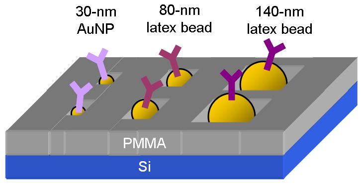
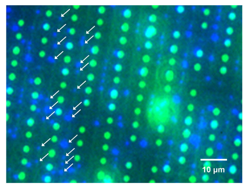
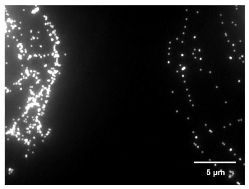
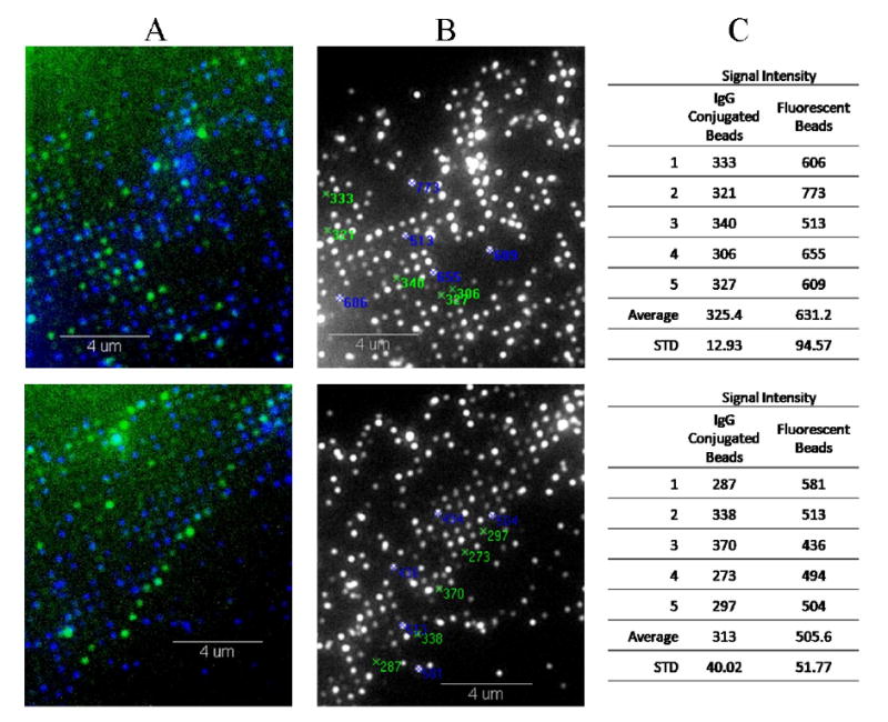
A protein nanoarray is created when submicro and nano beads, varying in their size and each conjugated with different proteins, self-assemble to specific locations depending on the diameter matching the surface electron beam patterns created. Protein binding is confirmed from the fluorescence attenuation of the beads upon antigen-antibody binding on the bead surface. This method, called size-dependent self-assembly, allows control of the location of each type of bead, and thus, control of the location of multiple proteins. It provides fast multi-component patterning with a high binding resolution, which can be detected using a fluorescent light microscope. This method is developed to be a simple stand-alone tool for analysis of protein interactions. In addition, it has the potential to be used in conjunction with other methods protein analysis methods, such as enzyme-linked immunosorbent assay (ELISA) and atomic force microscopy (AFM).
Figures
References
-
- Lee JH, Shim HW, Choi HS, Son YA, Lee CS. J Phys Chem Solids. 2008;69:1581–1584.
-
- Blawas AS, Reichert WM. Biomaterials. 1998;19:595–609. - PubMed
-
- Saravia V, Kupeu S, Nolte M, Huber C, Pum D, Fery A, Sleytr UB, Toca-Herrera JL. J Biotechnol. 2007;130:247–252. - PubMed
-
- Natarajan S, Katsamba PS, Miles A, Eckman J, Papalia GA, Rich RL, Gale BK, Myszka DG. Anal Biochem. 2008;373:141–146. - PubMed
-
- Lee KB, Park SJ, Mirkin CA, Smith JC, Mrksich M. Science. 2002;295:1702–1705. - PubMed
Grants and funding
LinkOut - more resources
Full Text Sources
Miscellaneous
