

Usefulness of hemodynamic sensors for physiologic cardiac pacing in heart failure patients
- PMID: 21461359
- PMCID: PMC3065053
- DOI: 10.4061/2011/925653
Usefulness of hemodynamic sensors for physiologic cardiac pacing in heart failure patients
Abstract
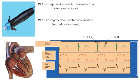
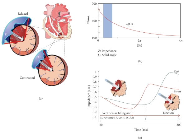
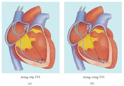
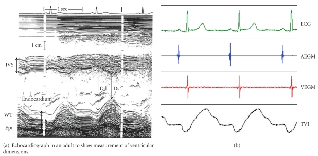
The rate adaptive sensors applied to cardiac pacing should respond as promptly as the normal sinus node with an highly specific and sensitive detection of the need of increasing heart rate. Sensors operating alone may not provide optimal heart responsiveness: central venous pH sensing, variations in the oxygen content of mixed venous blood, QT interval, breathing rate and pulmonary minute ventilation monitored by thoracic impedance variations, activity sensors. Using sensors that have different attributes but that work in a complementary manners offers distinct advantages. However, complicated sensors interactions may occur. Hemodynamic sensors detect changes in the hemodynamic performances of the heart, which partially depends on the autonomic nervous system-induced inotropic regulation of myocardial fibers. Specific hemodynamic sensors have been designed to measure different expression of the cardiac contraction strength: Peak Endocardial Acceleration (PEA), Closed Loop Stimulation (CLS) and TransValvular Impedance (TVI), guided by intraventricular impedance variations. Rate-responsive pacing is just one of the potential applications of hemodynamic sensors in implantable pacemakers. Other issues discussed in the paper include: hemodynamic monitoring for the optimal programmation and follow up of patients with cardiac resynchronization therapy; hemodynamic deterioration impact of tachyarrhythmias; hemodynamic upper rate limit control; monitoring and prevention of vasovagal malignant syncopes.
Figures
Similar articles
-
An implantable intracardiac accelerometer for monitoring myocardial contractility. The Multicenter PEA Study Group.Pacing Clin Electrophysiol. 1996 Dec;19(12 Pt 1):2066-71. doi: 10.1111/j.1540-8159.1996.tb03280.x. Pacing Clin Electrophysiol. 1996. PMID: 8994945
-
Is local myocardial contractility related to endocardial acceleration signals detected by a transvenous pacing lead?Pacing Clin Electrophysiol. 1996 Nov;19(11 Pt 2):1682-8. doi: 10.1111/j.1540-8159.1996.tb03206.x. Pacing Clin Electrophysiol. 1996. PMID: 8945024
-
The DDDR closed loop stimulation for the prevention of vasovagal syncope: results from the INVASY prospective feasibility registry.Europace. 2003 Apr;5(2):153-62. doi: 10.1053/eupc.2002.0292. Europace. 2003. PMID: 12633640
-
Sensors for rate-adaptive pacing: How they work, strengths, and limitations.J Cardiovasc Electrophysiol. 2020 Nov;31(11):3009-3027. doi: 10.1111/jce.14733. Epub 2020 Oct 8. J Cardiovasc Electrophysiol. 2020. PMID: 32877004 Review.
-
Rate Adaptive Pacing: Memories From a Bygone Era.Heart Lung Circ. 2021 Feb;30(2):225-232. doi: 10.1016/j.hlc.2020.08.023. Epub 2020 Oct 5. Heart Lung Circ. 2021. PMID: 33032894 Review.
Cited by
-
Rate-Responsive Cardiac Pacing: Technological Solutions and Their Applications.Sensors (Basel). 2023 Jan 27;23(3):1427. doi: 10.3390/s23031427. Sensors (Basel). 2023. PMID: 36772467 Free PMC article. Review.
-
Biological pacemaker created by minimally invasive somatic reprogramming in pigs with complete heart block.Sci Transl Med. 2014 Jul 16;6(245):245ra94. doi: 10.1126/scitranslmed.3008681. Sci Transl Med. 2014. PMID: 25031269 Free PMC article.
-
Chronotropic incompetence in Chagas disease: effectiveness of blended sensor (volume/minute and accelerometer).Rev Bras Cir Cardiovasc. 2015 Jul-Sep;30(3):311-5. doi: 10.5935/1678-9741.20150035. Rev Bras Cir Cardiovasc. 2015. PMID: 26313721 Free PMC article.
References
-
- Rickards AF, Donaldson RM. Rate-responsive pacing. Pacing and Clinical Electrophysiology. 1983;1:12–19. - PubMed
-
- Gwinn N, Leman R, Kratz J, White JK, Zile MR, Gillette P. Chronotropic incompetence: a common and progressive finding in pacemaker patients. American Heart Journal. 1992;123(5):1216–1219. - PubMed
-
- Leung SK, Lau CP, Tang MO. Cardiac output is a sensitive indicator of difference in exercise performance between single and dual sensor pacemakers. Pacing and Clinical Electrophysiology. 1998;21(1):35–41. - PubMed
-
- Karloef I. Haemodynamic effect of atrial triggered versus fixed rate pacing at rest and during exercise in complete heart block. Acta Medica Scandinavica. 1975;197(3):195–206. - PubMed
-
- Fananapazir L, Bennett DH, Monks P. Atrial synchronized ventricular pacing: contribution of the chronotropic response to improved exercise performance. Pacing and Clinical Electrophysiology. 1983;6(3):601–610. - PubMed
LinkOut - more resources
Full Text Sources