. 2011 Mar 15:2011:716871.
doi: 10.4061/2011/716871.

Mitochondrial dysfunction in Parkinson's disease

Affiliations

Mitochondrial dysfunction in Parkinson's disease

P C Keane et al. Parkinsons Dis. .

Abstract

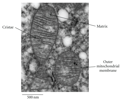
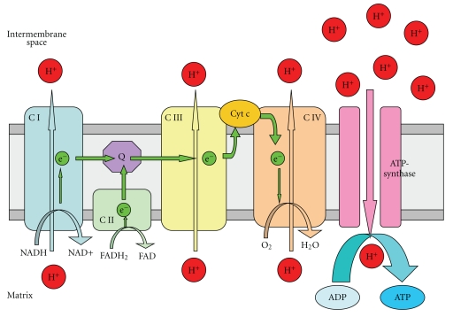
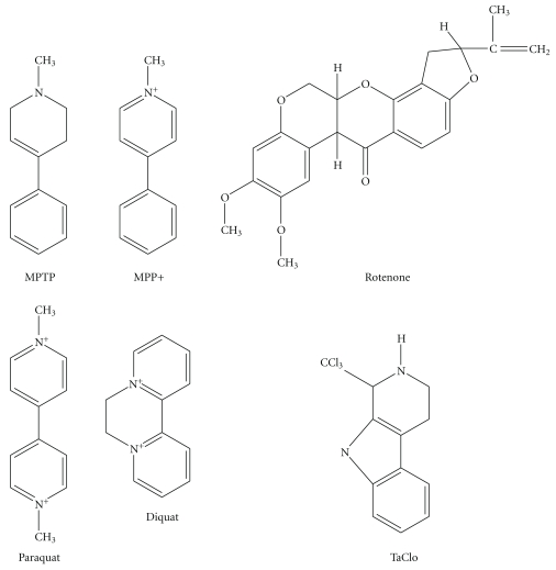
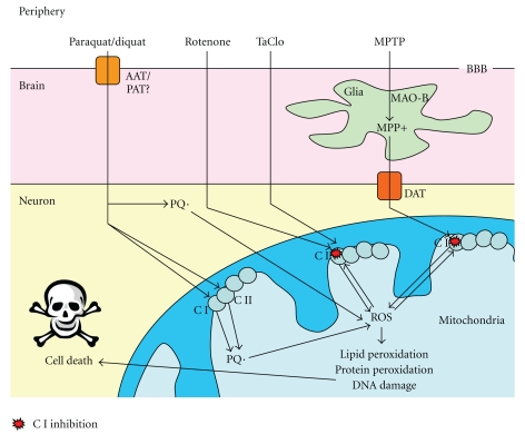
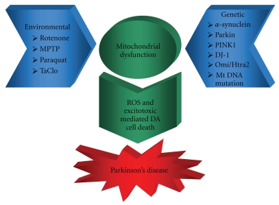
Parkinson's disease (PD) is a progressive, neurodegenerative condition that has increasingly been linked with mitochondrial dysfunction and inhibition of the electron transport chain. This inhibition leads to the generation of reactive oxygen species and depletion of cellular energy levels, which can consequently cause cellular damage and death mediated by oxidative stress and excitotoxicity. A number of genes that have been shown to have links with inherited forms of PD encode mitochondrial proteins or proteins implicated in mitochondrial dysfunction, supporting the central involvement of mitochondria in PD. This involvement is corroborated by reports that environmental toxins that inhibit the mitochondrial respiratory chain have been shown to be associated with PD. This paper aims to illustrate the considerable body of evidence linking mitochondrial dysfunction with neuronal cell death in the substantia nigra pars compacta (SNpc) of PD patients and to highlight the important need for further research in this area.


Figures

Figure 1
Figure 1
An electron micrograph to show mitochondrial ultrastructure. Reproduced with the kind permission of Tracy Davey, EM Research Services, Newcastle University.
Figure 2
Figure 2
Mitochondrial electron transport chain: schematic representation of the mitochondrial electron transport chain involved in oxidative phosphorylation. CI and II (Complexes I and II) transport electrons (es) generated by the conversion of NADH to NAD+ (CI) or FADH2 to FAD (CII) through Q (ubiquinone), CIII, Cyt c (cytochrome c) and finally CIV, which uses an e to convert O2 to H2O. During electron transfer, CI, II, and IV pump protons (H+s) from the mitochondrial matrix into the intermembrane space generating a H+ concentration gradient that drives the formation of ATP from ADP by ATP-synthase (Complex V).
Figure 3
Figure 3
Structures of the PD-linked neurotoxins MPTP/MPP+, Rotenone, Paraquat, Diquat, and TaClo.
Figure 4
Figure 4
Mechanisms of mitochondrial dysfunction-mediated cell death generated by neurotoxins: Paraquat crosses the blood brain barrier (BBB) by an as yet unclear mechanism, possibly via an amino acid transporter (AAT)/polyamine transporter (PAT), where it enters the cell and is spontaneously reduced to the paraquat radical PQ· or reduced by Complex I or II (CI or II) and can then form reactive oxygen species (ROS). Rotenone and TaClo enter neurons and can cause CI inhibition which leads to the production of ROS. MPTP enters the brain, where it is converted to MPP+ in glial cells by monoamine oxidase-B (MAO-B) which is transported into neurons by the dopamine transporter (DAT). Once in the cell, MPP+ also causes CI inhibition and generation of ROS. ROS formed by these environmental toxins can then exacerbate the CI inhibition as well as causing lipid and protein peroxidation, DNA damage, and, ultimately, cell death.
Figure 5
Figure 5
Schematic representation of mitochondrial dysfunction in Parkinson's disease: environmental and genetic factors combine to cause mitochondrial dysfunction leading to ROS- and excitotoxic mediated DA cell death and Parkinson's disease.

References

    1. Lees AJ, Hardy J, Revesz T. Parkinson’s disease. The Lancet. 2009;373(9680):2055–2066. - PubMed
    1. Dickson DW, Braak H, Duda JE, et al. Neuropathological assessment of Parkinson’s disease: refining the diagnostic criteria. The Lancet Neurology. 2009;8(12):1150–1157. - PubMed
    1. Keeney PM, Xie J, Capaldi RA, Bennett JP. Parkinson’s disease brain mitochondrial complex I has oxidatively damaged subunits and is functionally impaired and misassembled. Journal of Neuroscience. 2006;26(19):5256–5264. - PMC - PubMed
    1. Schapira AHV, Cooper JM, Dexter D, Clark JB, Jenner P, Marsden CD. Mitochondrial complex I deficiency in Parkinson’s disease. Journal of Neurochemistry. 1990;54(3):823–827. - PubMed
    1. Kitada T, Asakawa S, Hattori N, et al. Mutations in the parkin gene cause autosomal recessive juvenile parkinsonism. Nature. 1998;392(6676):605–608. - PubMed

LinkOut - more resources