

Characterization of the E506Q and H537A dysfunctional mutants in the E. coli ABC transporter MsbA
- PMID: 21462989
- PMCID: PMC3128438
- DOI: 10.1021/bi101666p
Characterization of the E506Q and H537A dysfunctional mutants in the E. coli ABC transporter MsbA
Abstract
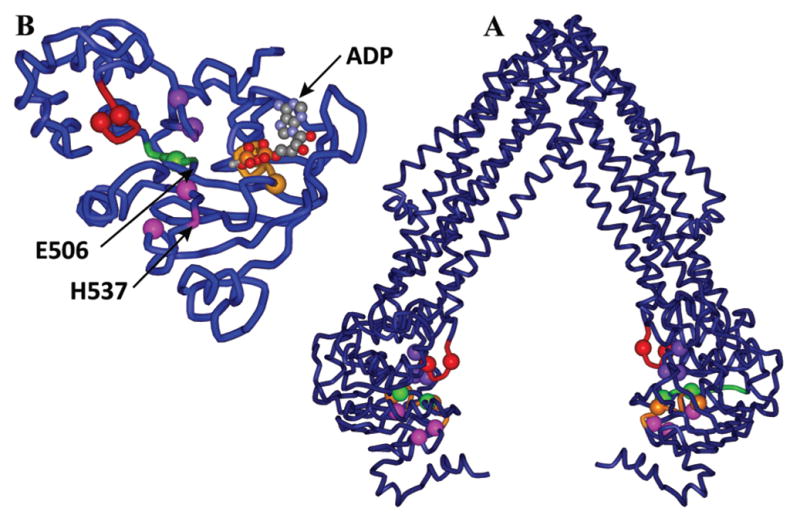
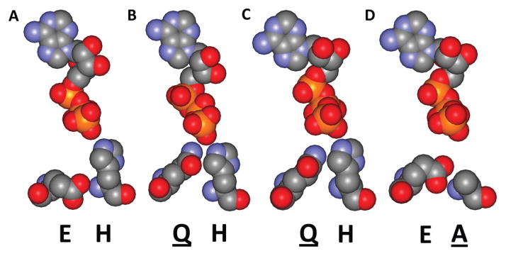
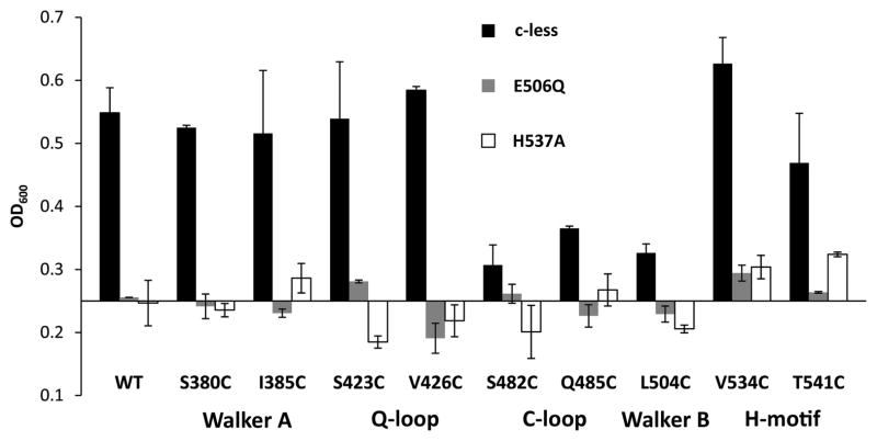
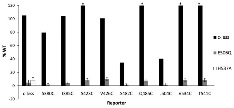
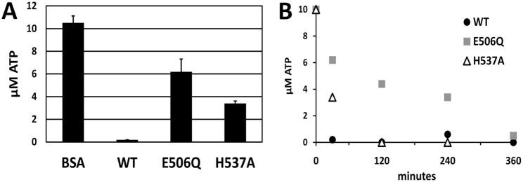
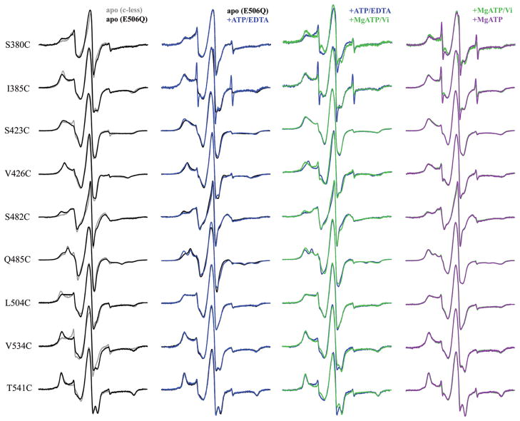
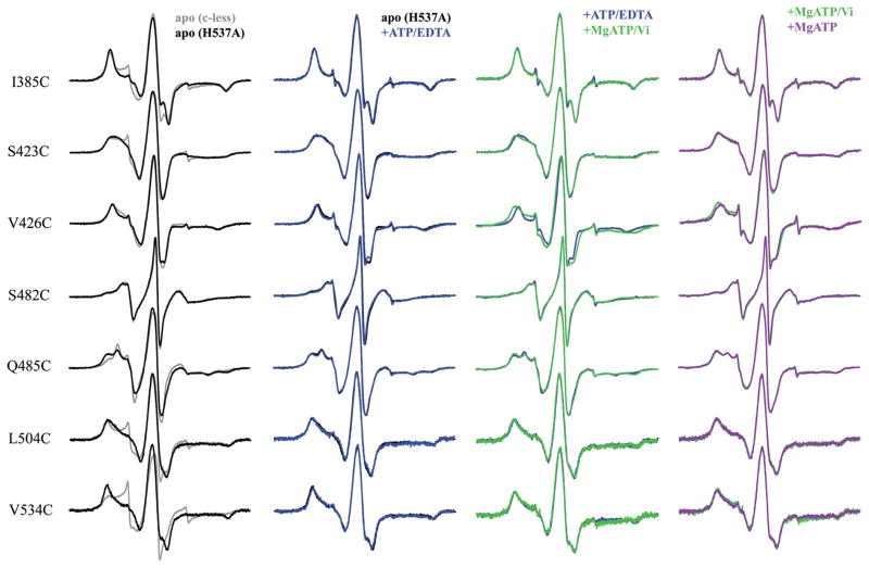
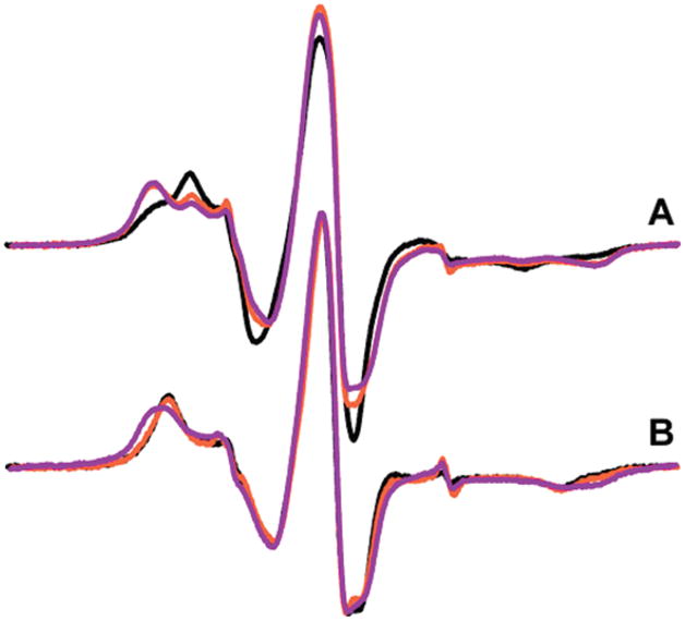
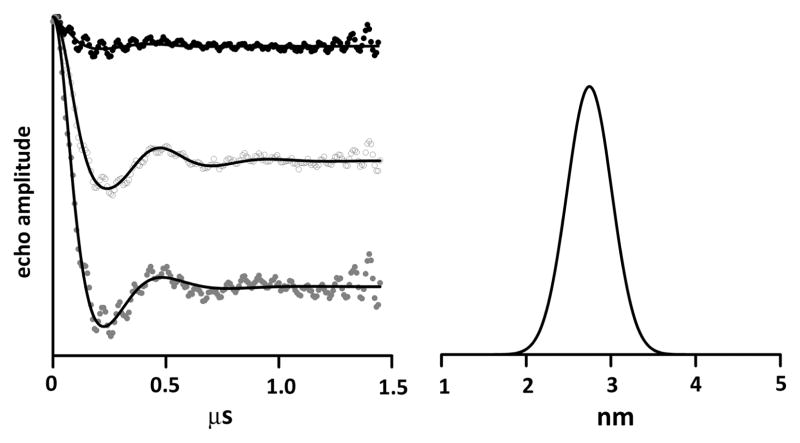
MsbA is a member of the ABC transporter superfamily that is specifically found in Gram-negative bacteria and is homologous to proteins involved in both bacterial and human drug resistance. The E506Q and H537A mutations have been introduced and used for crystallization of other members of the ABC transporter protein family, including BmrA and the ATPase domains MalK, HlyB-NBD, and MJ0796, but have not been previously studied in detail or investigated in the MsbA lipid A exporter. We utilized an array of biochemical and EPR spectroscopy approaches to characterize the local and global effects of these nucleotide binding domain mutations on the E. coli MsbA homodimer. The lack of cell viability in an in vivo growth assay confirms that the presence of the E506Q or H537A mutations within MsbA creates a dysfunctional protein. To further investigate the mode of dysfunction, a fluorescent ATP binding assay was used and showed that both mutant proteins maintain their ability to bind ATP, but ATPase assays indicate hydrolysis is severely inhibited by each mutation. EPR spectroscopy data using previously identified and characterized reporter sites within the nucleotide binding domain along with ATP detection assays show that hydrolysis does occur over time in both mutants, though more readily in the H537A protein. DEER spectroscopy demonstrates that both proteins studied are purified in a closed dimer conformation, indicating that events within the cell can induce a stable, closed conformation of the MsbA homodimer that does not reopen even in the absence of nucleotide.
Figures
Similar articles
-
Effects of the L511P and D512G mutations on the Escherichia coli ABC transporter MsbA.Biochemistry. 2011 Apr 5;50(13):2594-602. doi: 10.1021/bi1018418. Epub 2011 Mar 8. Biochemistry. 2011. PMID: 21344946 Free PMC article.
-
Asymmetry in the homodimeric ABC transporter MsbA recognized by a DARPin.J Biol Chem. 2012 Jun 8;287(24):20395-406. doi: 10.1074/jbc.M112.359794. Epub 2012 Apr 20. J Biol Chem. 2012. PMID: 22523072 Free PMC article.
-
Resting state conformation of the MsbA homodimer as studied by site-directed spin labeling.Biochemistry. 2004 Jul 6;43(26):8600-6. doi: 10.1021/bi0497751. Biochemistry. 2004. PMID: 15222771
-
Investigating the dynamic nature of the ABC transporters: ABCB1 and MsbA as examples for the potential synergies of MD theory and EPR applications.Biochem Soc Trans. 2015 Oct;43(5):1023-32. doi: 10.1042/BST20150138. Biochem Soc Trans. 2015. PMID: 26517918 Review.
-
MsbA: an ABC transporter paradigm.Biochem Soc Trans. 2021 Dec 17;49(6):2917-2927. doi: 10.1042/BST20211030. Biochem Soc Trans. 2021. PMID: 34821931 Review.
Cited by
-
Mapping Free Energy Pathways for ATP Hydrolysis in the E. coli ABC Transporter HlyB by the String Method.Molecules. 2018 Oct 16;23(10):2652. doi: 10.3390/molecules23102652. Molecules. 2018. PMID: 30332773 Free PMC article.
-
The deviant ATP-binding site of the multidrug efflux pump Pdr5 plays an active role in the transport cycle.J Biol Chem. 2013 Oct 18;288(42):30420-30431. doi: 10.1074/jbc.M113.494682. Epub 2013 Sep 9. J Biol Chem. 2013. PMID: 24019526 Free PMC article.
-
Locating a lipid at the portal to the lipoxygenase active site.Biophys J. 2012 Nov 21;103(10):2134-44. doi: 10.1016/j.bpj.2012.10.002. Epub 2012 Nov 20. Biophys J. 2012. PMID: 23200047 Free PMC article.
-
ATP-Binding Cassette Transporter Structure Changes Detected by Intramolecular Fluorescence Energy Transfer for High-Throughput Screening.Mol Pharmacol. 2015 Jul;88(1):84-94. doi: 10.1124/mol.114.096792. Epub 2015 Apr 29. Mol Pharmacol. 2015. PMID: 25924616 Free PMC article.
-
Mechanistic picture for conformational transition of a membrane transporter at atomic resolution.Proc Natl Acad Sci U S A. 2013 Nov 19;110(47):18916-21. doi: 10.1073/pnas.1313202110. Epub 2013 Nov 4. Proc Natl Acad Sci U S A. 2013. PMID: 24191018 Free PMC article.
References
-
- Cotten JF, Welsh MJ. Covalent modification of the regulatory domain irreversibly stimulates cystic fibrosis transmembrane conductance regulator. J Biol Chem. 1997;272:25617–25622. - PubMed
-
- Allikmets R, Singh N, Sun H, Shroyer NF, Hutchinson A, Chidambaram A, Gerrard B, Baird L, Stauffer D, Peiffer A, Rattner A, Smallwood P, Li Y, Anderson KL, Lewis RA, Nathans J, Leppert M, Dean M, Lupski JR. A photoreceptor cell-specific ATP-binding transporter gene (ABCR) is mutated in recessive Stargardt macular dystrophy. Nature Genet. 1997;15:236–246. - PubMed
-
- Karow M, Georgopoulos C. The essential Escherichia coli msbA gene, a multicopy suppressor of null mutations in the htrB gene, is related to the universally conserved family of ATP-dependent translocators. Mol Microbiol. 1993;7:69–79. - PubMed
Publication types
MeSH terms
Substances
Grants and funding
LinkOut - more resources
Full Text Sources
Molecular Biology Databases
