

Imipramine treatment improves cognitive outcome associated with enhanced hippocampal neurogenesis after traumatic brain injury in mice
- PMID: 21463148
- PMCID: PMC3113418
- DOI: 10.1089/neu.2010.1563
Imipramine treatment improves cognitive outcome associated with enhanced hippocampal neurogenesis after traumatic brain injury in mice
Abstract
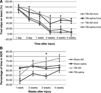
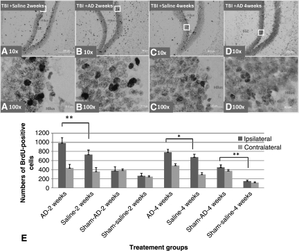
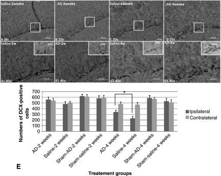
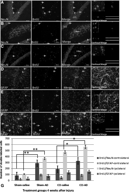
Previous animal and human studies have demonstrated that chronic treatment with several different antidepressants can stimulate neurogenesis, neural remodeling, and synaptic plasticity in the normal hippocampus. Imipramine is a commonly used tricyclic antidepressant (TCA). We employed a controlled cortical impact (CCI) mouse model of traumatic brain injury (TBI) to assess the effect of imipramine on neurogenesis and cognitive and motor function recovery after TBI. Mice were given daily imipramine injections for either 2 or 4 weeks after injury. Bromodeoxyuridine (BrdU) was administered 3-7 days post-brain injury to label the cells that proliferated as a result of the injury. We assessed the effects of imipramine on post-traumatic motor function using a beam-walk test and an assessment of cognitive function: the novel object recognition test (NOR). Histological analyses were performed at 2 and 4 weeks after CCI. Brain-injured mice treated with imipramine showed significantly improved cognitive function compared to a saline-treated group (p<0.001). However, there was no significant difference in motor function recovery between imipramine-treated and saline-treated mice. Histological examination revealed increased preservation of proliferation of Ki-67- and BrdU-positive cells in the hippocampal dentate gyrus (DG) at 2 and 4 weeks after TBI. Immunofluorescence double-labeling with BrdU and neuron-specific markers at 4 weeks after injury showed that most progenitors became neurons in the DG and astrocytes in the hilus. Notably, treatment with imipramine increased preservation of the total number of newly-generated neurons. Our findings provide direct evidence that imipramine treatment contributes to cognitive improvement after TBI, perhaps by enhanced hippocampal neurogenesis.
Figures

Similar articles
-
Basic fibroblast growth factor-enhanced neurogenesis contributes to cognitive recovery in rats following traumatic brain injury.Exp Neurol. 2009 Mar;216(1):56-65. doi: 10.1016/j.expneurol.2008.11.011. Epub 2008 Nov 27. Exp Neurol. 2009. PMID: 19100261 Free PMC article.
-
Imipramine treatment increases cell proliferation following fluid percussion brain injury in rats.Neurol Res. 2013 Apr;35(3):247-54. doi: 10.1179/1743132813Y.0000000164. Neurol Res. 2013. PMID: 23485052
-
Impact of inhibition of erythropoietin treatment-mediated neurogenesis in the dentate gyrus of the hippocampus on restoration of spatial learning after traumatic brain injury.Exp Neurol. 2012 May;235(1):336-44. doi: 10.1016/j.expneurol.2012.02.015. Epub 2012 Mar 4. Exp Neurol. 2012. PMID: 22414310 Free PMC article.
-
Hippocampal injury-induced cognitive and mood dysfunction, altered neurogenesis, and epilepsy: can early neural stem cell grafting intervention provide protection?Epilepsy Behav. 2014 Sep;38:117-24. doi: 10.1016/j.yebeh.2013.12.001. Epub 2014 Jan 13. Epilepsy Behav. 2014. PMID: 24433836 Free PMC article. Review.
-
Potential Role of Adult Hippocampal Neurogenesis in Traumatic Brain Injury.Curr Med Chem. 2022;29(19):3392-3419. doi: 10.2174/0929867328666210923143713. Curr Med Chem. 2022. PMID: 34561977 Review.
Cited by
-
Posttraining Epinephrine Reverses Memory Deficits Produced by Traumatic Brain Injury in Rats.Scientifica (Cairo). 2016;2016:9151490. doi: 10.1155/2016/9151490. Epub 2016 Apr 4. Scientifica (Cairo). 2016. PMID: 27127685 Free PMC article.
-
Endogenous neurogenic cell response in the mature mammalian brain following traumatic injury.Exp Neurol. 2016 Jan;275 Pt 3(0 3):405-410. doi: 10.1016/j.expneurol.2015.04.017. Epub 2015 Apr 30. Exp Neurol. 2016. PMID: 25936874 Free PMC article. Review.
-
Expression of monoamine transporters, nitric oxide synthase 3, and neurotrophin genes in antidepressant-stimulated astrocytes.Front Psychiatry. 2012 Apr 20;3:33. doi: 10.3389/fpsyt.2012.00033. eCollection 2012. Front Psychiatry. 2012. PMID: 22529824 Free PMC article.
-
Hippocampal BMP signaling as a common pathway for antidepressant action.Cell Mol Life Sci. 2021 Dec 22;79(1):31. doi: 10.1007/s00018-021-04026-y. Cell Mol Life Sci. 2021. PMID: 34936033 Free PMC article.
-
The Controlled Cortical Impact Model of Experimental Brain Trauma: Overview, Research Applications, and Protocol.Methods Mol Biol. 2016;1462:177-92. doi: 10.1007/978-1-4939-3816-2_11. Methods Mol Biol. 2016. PMID: 27604719 Free PMC article. Review.
References
-
- Ariza M. Serra-Grabulosa J.M. Junque C. Ramirez B. Mataro M. Poca A. Bargallo N. Sahuquillo J. Hippocampal head atrophy after traumatic brain injury. Neuropsychologia. 2006;44:1956–1961. - PubMed
-
- Bessa J.M. Ferreira D. Melo I. Marques F. Cerqueira J.J. Palha J.A. Almeida O.F. Sousa N. The mood-improving actions of antidepressants do not depend on neurogenesis but are associated with neuronal remodeling. Mol. Psychiatry. 2009;14:764–773. 739. - PubMed
-
- Bhattacharjee Y. Neuroscience. Shell shock revisited: solving the puzzle of blast trauma. Science. 2008;319:406–408. - PubMed
Publication types
MeSH terms
Substances
Grants and funding
LinkOut - more resources
Full Text Sources