

Irinotecan-induced alterations in intestinal cell kinetics and extracellular matrix component expression in the Dark Agouti rat
- PMID: 21463374
- PMCID: PMC3193150
- DOI: 10.1111/j.1365-2613.2011.00771.x
Irinotecan-induced alterations in intestinal cell kinetics and extracellular matrix component expression in the Dark Agouti rat
Abstract
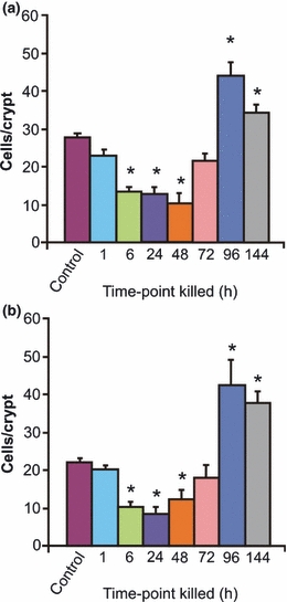
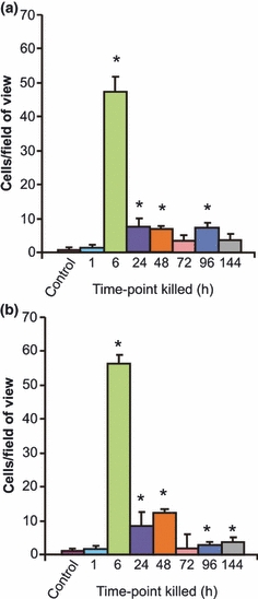
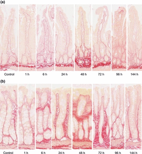
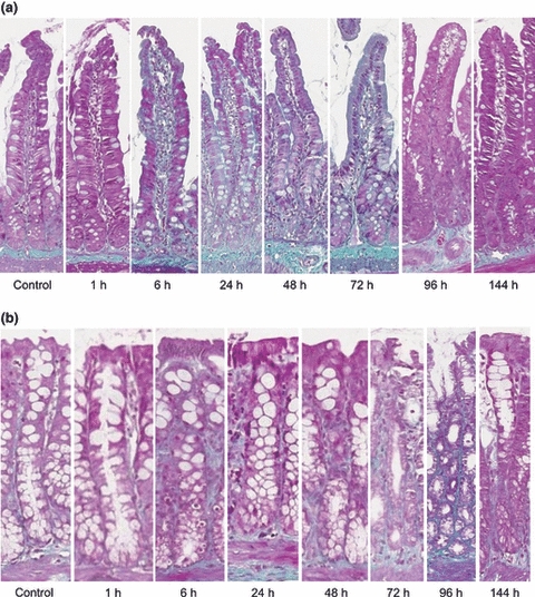
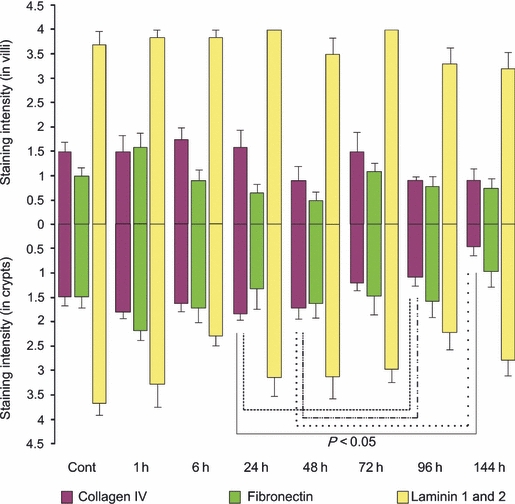
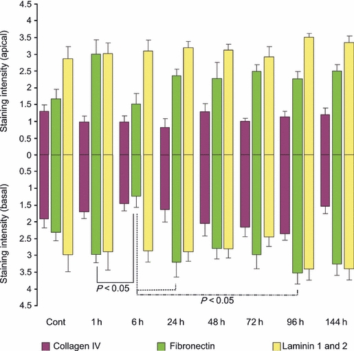
Chemotherapy-induced mucositis is characterized by damage of mucous membranes throughout the alimentary tract (AT). Extracellular matrix (ECM) components play a vital role in maintaining mucosal barrier integrity by regulating cellular apoptosis, proliferation and differentiation of overlying epithelial cells. The aims of this study were to characterize the changes in epithelial cell kinetics and to investigate the expression of the ECM components in the gastrointestinal tract following irinotecan administration. Female dark agouti rats were treated with single 200 mg/kg dose irinotecan and killed at various time points (1, 6, 24, 48, 72, 96 and 14 h) after treatment. Ki67 immunostaining and TUNEL were used to assess proliferation and apoptosis, respectively, in the jejunum and colon. Masson trichrome staining and picro-sirius red staining were used to determine the level of collagen, and immunohistochemistry was used to further assess collagen IV, fibronectin and laminin 1 and 2 expression in these tissues. Irinotecan halved cellular proliferation in the jejunum and colon at 48 and 24 h, respectively, while apoptosis peaked at 6 h (P < 0.05). There was a substantial increase in total collagen deposits around crypts from 24 h in both regions. However, collagen IV expression decreased significantly in the crypt region in a delayed fashion (P < 0.05). Fibronectin expression decreased significantly in jejunum and colon from 6 to 24 h following treatment (P < 0.05). Irinotecan induced a significant alteration in epithelial cell kinetics in both the jejunum and colon, and this correlated with changes in ECM component expression. Changes in ECM expression may have a direct impact on the loss of mucosal layer integrity evident in chemotherapy-induced mucositis.
© 2011 The Authors. International Journal of Experimental Pathology © 2011 International Journal of Experimental Pathology.
Figures
References
-
- Al-Dasooqi N, Gibson R, Bowen J, et al. Matrix metalloproteinases are possible mediators for the development of alimentary tract mucositis in the DA rat. Exp. Biol. Med. 2010;235:1244–1256. - PubMed
-
- Beaulieu J. Extracellular matrix components and integrins in relationship to human intestinal epithelial cell differentiation. Prog. Histochem. Cytochem. 1997;31:1–78. - PubMed
-
- Bowen J, Gibson R, Keefe D, Cummins A. Cytotoxic chemotherapy up-regulates pro-apoptotic Bax and Bak in the small intestine of rats and humans. Pathology. 2005;37:56–62. - PubMed
-
- Gibson R, Bowen J, Inglis M, et al. Irinotecan causes severe small intestinal damage, as well as colonic damage, in the rat with implanted breast cancer. J. Gastroenterol. Hepatol. 2003;18:1095–1100. - PubMed
Publication types
MeSH terms
Substances
LinkOut - more resources
Full Text Sources
