. 2011 Apr 6;100(7):1608-16.
doi: 10.1016/j.bpj.2011.02.020.

Flagella and pili-mediated near-surface single-cell motility mechanisms in P. aeruginosa

Affiliations

Flagella and pili-mediated near-surface single-cell motility mechanisms in P. aeruginosa

Jacinta C Conrad et al. Biophys J. .

Abstract

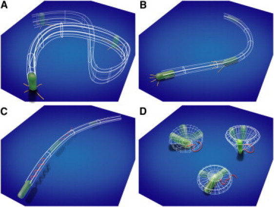
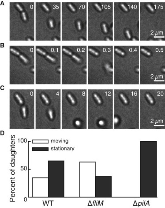
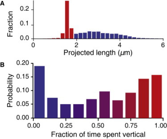
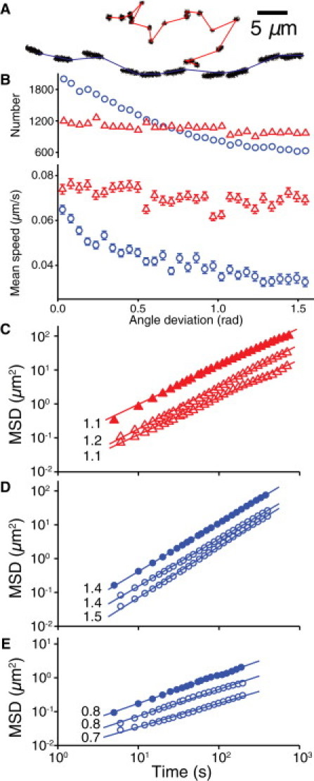
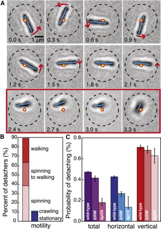
Bacterial biofilms are structured multicellular communities that are responsible for a broad range of infections. Knowing how free-swimming bacteria adapt their motility mechanisms near a surface is crucial for understanding the transition from the planktonic to the biofilm phenotype. By translating microscopy movies into searchable databases of bacterial behavior and developing image-based search engines, we were able to identify fundamental appendage-specific mechanisms for the surface motility of Pseudomonas aeruginosa. Type IV pili mediate two surface motility mechanisms: horizontally oriented crawling, by which the bacterium moves lengthwise with high directional persistence, and vertically oriented walking, by which the bacterium moves with low directional persistence and high instantaneous velocity, allowing it to rapidly explore microenvironments. The flagellum mediates two additional motility mechanisms: near-surface swimming and surface-anchored spinning, which often precedes detachment from a surface. Flagella and pili interact cooperatively in a launch sequence whereby bacteria change orientation from horizontal to vertical and then detach. Vertical orientation facilitates detachment from surfaces and thereby influences biofilm morphology.


Figures

Figure 1
Figure 1
Surface motility mechanisms observed for P. aeruginosa: TFP-driven (A) vertical walking and (B) horizontal crawling, and flagellum-driven (C) near-surface swimming and (D) surface-bound spinning.
Figure 2
Figure 2
TFP drive postdivision motility in P. aeruginosa. (A–C) Representative time series micrographs of postdivision behavior of daughter cells, showing (A) crawling, (B) detachment, and (C) walking. Timestamps in seconds are shown. (D) Percentage of daughter cells that exhibit motion or remain stationary after division, for the WT strain (N = 214 divisions), ΔfliM strain (N = 105 divisions), and ΔpilA strain (N = 131 divisions). No ΔpilA daughter cells move after division, indicating that postdivision surface motility must depend on TFP.
Figure 3
Figure 3
(A) Histogram of projected length and (B) probability of time spent oriented vertically for ΔfliM bacteria (N = 70,073 individual bacteria images).
Figure 4
Figure 4
Motility characteristics of the ΔfliM strain. (A) Representative trajectories of walking (top) and crawling (bottom) motility mechanisms, showing morphological differences. (B) Number and mean speed of walking (▵) and crawling (○) bacteria versus angle deviation for ΔfliM bacteria (N = 70,073). Error bars indicate 1 standard deviation (SD). (C) MSD versus time for walking bacteria over a 1-h measurement (▴) and density-limited 4- and 7-h measurements (▵). (D) MSD versus time for superdiffusive crawling bacteria for 1-h (●) and 4- and 7-h (○) measurements. (E) MSD versus time for subdiffusive trapped bacteria for 1-h (●) and 4- and 7-h (○) measurements.
Figure 5
Figure 5
(A) Histogram of projected length and (B) probability of time spent oriented vertically for WT bacteria (N = 170,073 individual bacteria images).
Figure 6
Figure 6
Motility characteristics of the WT strain. (A) Number and mean speed of walking (▵) and crawling (○) bacteria versus angle deviation for WT bacteria (N = 170,073). Error bars indicate 1 SD. (B) MSD versus time for walking (▴) and crawling (●) WT bacteria (N = 170,073); the dotted line indicates a slope of one (diffusive motion). Characteristic slopes match those of ΔfliM bacteria (33).
Figure 7
Figure 7
(A) Histogram of projected length for ΔpilA bacteria (N = 17,437 individual bacteria images). (B) MSD versus time for vertical (▴) and horizontal (●) ΔpilA bacteria (N = 17,437); the dotted line indicates a slope of 1 (diffusive motion).
Figure 8
Figure 8
Flagella-driven swimming and spinning motility. (A) Representative trajectories of swimming and spinning motility mechanisms. (B) Two-dimensional histogram of trajectory curvature and instantaneous velocity for swimming WT bacteria. Curvatures are calculated from three consecutive points in each trajectory. (C) Histogram of angular velocity for a representative spinning WT bacterium; positive angular velocity indicates clockwise motion.
Figure 9
Figure 9
Vertical orientation and appendage cooperation facilitate detachment. (A) Representative image series of a spinning WT bacterium detaching from the surface. Dots indicate the original center of rotation, dashed lines indicate the initial radius of the trajectory, solid lines indicate the bacterial backbone, and arrows indicate the direction and magnitude of rotation between consecutive images. Images inside the box outline (2.4–3.3 s) are those in which the bacterium has tilted off the surface. The bacterium rotates (0–0.9 s), slows (1.2–2.1 s), tilts away from the surface (2.4 s), and then detaches (3.3 s), using both flagella and TFP. (B) Percentage of detaching WT bacteria that exhibit out-of-plane (left) and in-plane (right) motility mechanisms. (C) Detachment probabilities for total, horizontal, and vertical bacteria as a function of strain (ΔfliM (N = 70,073), ΔpilA (N = 17,437), and WT (N = 170,073)). The WT consistently exhibit higher detachment probabilities, showing that both flagella and TFP facilitate detachment. Error bars indicate 1 SD.
Figure 10
Figure 10
Motility defects influence biofilm morphology. (A) Percentage of bacteria that exhibit the spinning motility mechanism or vertical orientation for ΔpilA (left, N = 376 and 270 at 0 h and 5.5 h) and WT (right, N = 355 and 257 at 0 h and 5.5 h) bacteria. Error bars indicate 1 SD. The percentage of spinning bacteria increases with time for ΔpilA because they cannot tilt up and detach. (B) Representative micrographs of ΔpilA (left), WT (right), and ΔfliM (right, inset) biofilms 5.5 h after inoculation. The presence of clusters in the ΔpilA biofilm compared with the uniform WT biofilm indicates that only TFP-competent bacteria are able to redistribute and detach. The ΔfliM strain, which has a small detachment defect and no motility defect, is nonuniform but contains no large clusters.

References

    1. Costerton J.W., Stewart P.S., Greenberg E.P. Bacterial biofilms: a common cause of persistent infections. Science. 1999;284:1318–1322. - PubMed
    1. Touhami A., Jericho M.H., Beveridge T.J. Nanoscale characterization and determination of adhesion forces of Pseudomonas aeruginosa pili by using atomic force microscopy. J. Bacteriol. 2006;188:370–377. - PMC - PubMed
    1. Anderson G.G., O'Toole G.A. Innate and induced resistance mechanisms of bacterial biofilms. Curr. Top. Microbiol. Immunol. 2008;322:85–105. - PubMed
    1. Hall-Stoodley L., Stoodley P. Evolving concepts in biofilm infections. Cell. Microbiol. 2009;11:1034–1043. - PubMed
    1. Nassif X., Marceau M., Taha M.K. Type-4 pili and meningococcal adhesiveness. Gene. 1997;192:149–153. - PubMed

Publication types

LinkOut - more resources