. 2011 Apr 6;100(7):1708-17.
doi: 10.1016/j.bpj.2011.02.039.

Activation of F-actin binding capacity of ezrin: synergism of PIP₂ interaction and phosphorylation

Affiliations

Activation of F-actin binding capacity of ezrin: synergism of PIP₂ interaction and phosphorylation

Sabine Bosk et al. Biophys J. .

Abstract

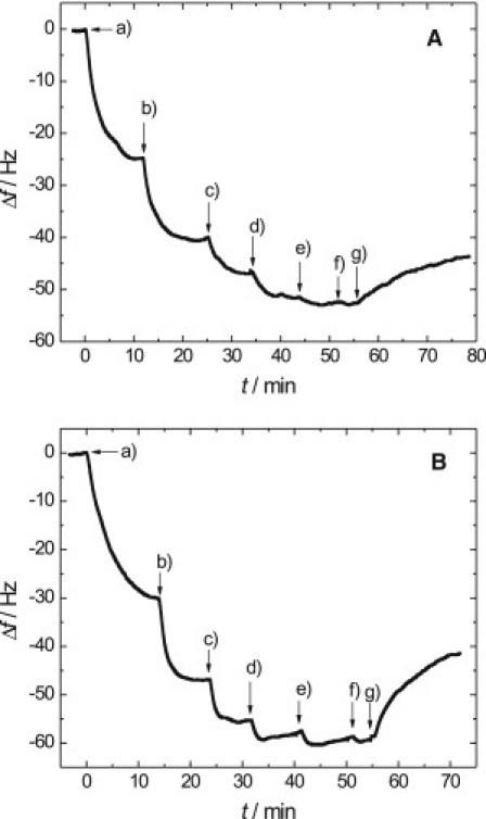
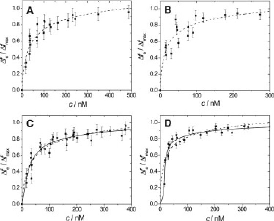
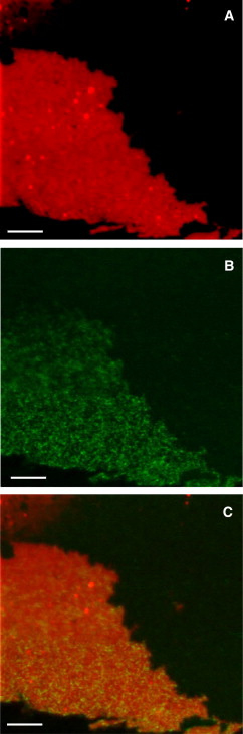
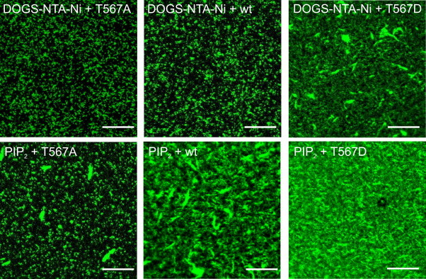
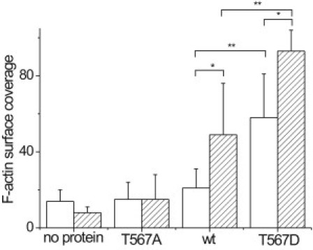
Ezrin is a membrane-cytoskeleton linker protein that can bind F-actin in its active conformation. Several means of regulation of ezrin's activity have been described including phosphorylation of Thr-567 and binding of L-α-phosphatidylinositol-4,5-bisphosphate (PIP(2)). However, the relative contributions of these events toward activation of the protein and their potential interdependence are not known. We developed an assay based on solid-supported membranes, to which different ezrin mutants (ezrin T567A (inactive mutant), wild-type, and T567D (active pseudophosphorylated mutant)) were bound, that enabled us to analyze the influence of phosphorylation and PIP(2) binding on ezrin's activation state in vitro. The lipid bilayers employed contained either DOGS-NTA-Ni to bind the proteins via an N-terminal His-tag, or PIP(2), to which ezrin binds via specific binding sites located in the N-terminal region of the protein. Quantitative analysis of the binding behavior of all three proteins to the two different receptor lipids revealed that all three bind with high affinity and specificity to the two receptor lipids. Fluorescence microscopy on ezrin-decorated solid-supported membranes showed that, dependent on the mode of binding and the phosphorylation state, ezrin is capable of binding actin filaments. A clear synergism between phosphorylation and the receptor lipid PIP(2) was observed, suggesting a conformational switch from the dormant to the active, F-actin binding state by recognition of PIP(2), which is enhanced by the phosphorylation.


Figures

Figure 1
Figure 1
Representative time course of the frequency shift upon stepwise ezrin wt addition (af) to a solid-supported (A) OT-DOPC/DOGS-NTA-Ni (9:1) or (B) OT-POPC/PIP2 (9:1) membrane in 20 mM Tris/HCl, 50 mM KCl, 0.1 mM EDTA, pH = 7.4. (g) Rinsing with pure buffer. Concentrations in panel A: (a) 13 nM, (b) 39 nM, (c) 90 nM, (d) 161 nM, (e) 249 nM, and (f) 280 nM. Concentrations in panel B: (a) 19 nM, (b) 44 nM, (c) 91 nM, (d) 148 nM, (e) 233 nM, and (f) 282 nM.
Figure 2
Figure 2
Adsorption isotherms of (A) ezrin T567A and (B) ezrin T567D binding to a DOGS-NTA-Ni-doped (10 mol %) SSM. Adsorption isotherms of (C) ezrin T567A and (D) ezrin T567D binding to a PIP2-doped (10 mol %) SSM. All experiments were performed in 20 mM Tris/HCl, 50 mM KCl, and 0.1 mM EDTA, pH = 7.4. (Dashed lines) Results of fitting Eq. 1 to the data. The results are summarized in Table 2. (Solid lines) Results of fitting Eq. 3 to the data. For further details, see text.
Figure 3
Figure 3
Confocal laser scanning fluorescence microscopy image of (A) AlexaFluor633-maleimide-labeled ezrin wt immobilized on a SSM containing 0.5% PIP2, 1% perylene, 98.5% POPC in F-buffer (20 mM Tris/HCl, 50 mM KCl, 2 mM MgCl2, and 2 mM ATP, pH = 7.4). (B) AlexaFluor488-phalloidin-labeled actin. (C) Overlay of the two images. Scale bars: 5 μm.
Figure 4
Figure 4
Confocal laser scanning fluorescence microscopy images of AlexaFluor488-phalloidin-labeled actin bound to ezrin on DOGS-NTA-Ni (10%) doped SSMs or PIP2 (10%) doped SSMs on silicon substrates using ezrin T567A, ezrin wt, and ezrin T567D as indicated. Scale bars: 10 μm.
Figure 5
Figure 5
Quantification of the surface coverage via pixel analysis using the program ImageJ (National Institutes of Health). Results obtained for DOGS-NTA-Ni (10 mol %) (open bars) and PIP2 (10 mol %) (hatched bars) doped SSMs on silicon substrates using ezrin and the mutants. These data were generated from at least 2–3 independently prepared samples, from which 3–7 images were taken at different regions of the surface for coverage determination. The values are given as mean values ± SD. A Wilcoxon rank-sum test confirms that the difference in F-actin coverage is significantly different for ezrin wt bound to DOGS-NTA-Ni and PIP2 (P = 0.0007, ) and ezrin T567D bound to DOGS-NTA-Ni and PIP2 (P = 0.007, ). In addition, the differences in F-actin coverage comparing ezrin wt and ezrin T567D bound to DOGS-NTA-Ni (P = 0.00003, ∗∗) and ezrin wt and ezrin T567D bound to PIP2 (P = 0.0004, ∗∗) showed high significance.

Similar articles

Cited by

References

    1. Fehon R.G., McClatchey A.I., Bretscher A. Organizing the cell cortex: the role of ERM proteins. Nat. Rev. Mol. Cell Biol. 2010;11:276–287. - PMC - PubMed
    1. Gary R., Bretscher A. Ezrin self-association involves binding of an N-terminal domain to a normally masked C-terminal domain that includes the F-actin binding site. Mol. Biol. Cell. 1995;6:1061–1075. - PMC - PubMed
    1. Zhu L.X., Hatakeyama J., Forte J.G. Comparative study of ezrin phosphorylation among different tissues: more is good; too much is bad. Am. J. Physiol. Cell Physiol. 2008;295:C192–C202. - PMC - PubMed
    1. Turunen O., Wahlström T., Vaheri A. Ezrin has a COOH-terminal actin-binding site that is conserved in the ezrin protein family. J. Cell Biol. 1994;126:1445–1453. - PMC - PubMed
    1. Nakamura F., Huang L., Furthmayr H. Regulation of F-actin binding to platelet moesin in vitro by both phosphorylation of threonine 558 and polyphosphatidylinositides. Mol. Biol. Cell. 1999;10:2669–2685. - PMC - PubMed

Publication types

LinkOut - more resources