

Study of cell-matrix adhesion dynamics using surface plasmon resonance imaging ellipsometry
- PMID: 21463596
- PMCID: PMC3072622
- DOI: 10.1016/j.bpj.2011.01.033
Study of cell-matrix adhesion dynamics using surface plasmon resonance imaging ellipsometry
Abstract
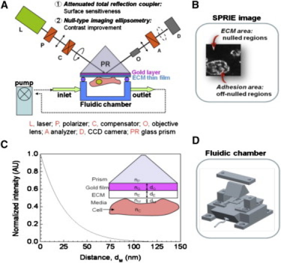
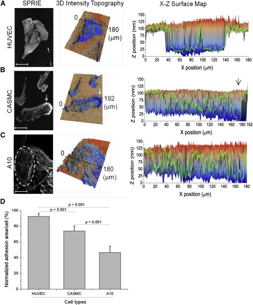
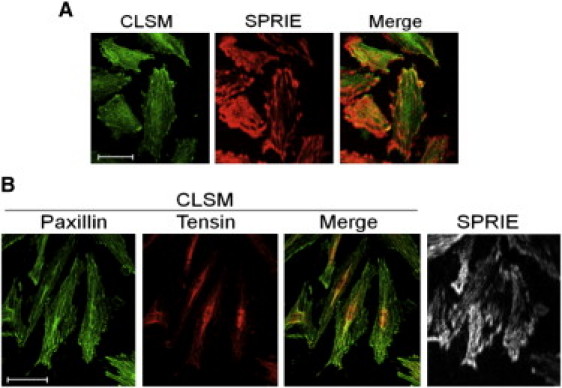
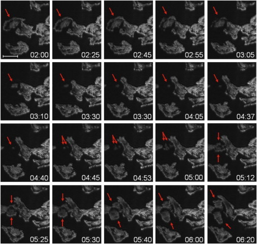
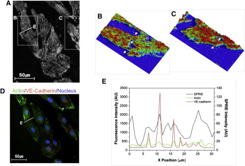
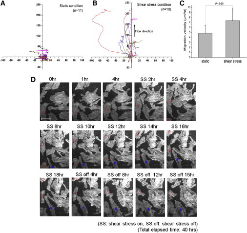
The interaction of cells with extracellular matrix, termed cell-matrix adhesions, importantly governs multiple cellular phenomena. Knowledge of the functional dynamics of cell-matrix adhesion could provide critical clues for understanding biological phenomena. We developed surface plasmon resonance imaging ellipsometry (SPRIE) to provide high contrast images of the cell-matrix interface in unlabeled living cells. To improve the contrast and sensitivity, the null-type imaging ellipsometry technique was integrated with an attenuated total reflection coupler. We verified that the imaged area of SPRIE was indeed a cell-matrix adhesion area by confocal microscopy imaging. Using SPRIE, we demonstrated that three different cell types exhibit distinct features of adhesion. SPRIE was applied to diverse biological systems, including during cell division, cell migration, and cell-cell communication. We imaged the cell-matrix anchorage of mitotic cells, providing the first label-free imaging of this interaction to our knowledge. We found that cell-cell communication can alter cell-matrix adhesion, possibly providing direct experimental evidence for cell-cell communication-mediated changes in cell adhesion. We also investigated shear-stress-induced adhesion dynamics in real time. Based on these data, we expect that SPRIE will be a useful methodology for studying the role of cell-matrix adhesion in important biological phenomena.
Copyright © 2011 Biophysical Society. Published by Elsevier Inc. All rights reserved.
Figures
Similar articles
-
Recent advances in label-free imaging of cell-matrix adhesions.Chem Commun (Camb). 2023 Feb 23;59(17):2341-2351. doi: 10.1039/d2cc06499e. Chem Commun (Camb). 2023. PMID: 36744880 Review.
-
The control of endothelial cell adhesion and migration by shear stress and matrix-substrate anchorage.Biomaterials. 2012 Mar;33(7):1959-69. doi: 10.1016/j.biomaterials.2011.11.017. Epub 2011 Dec 10. Biomaterials. 2012. PMID: 22154622
-
Mapping the dynamics of force transduction at cell-cell junctions of epithelial clusters.Elife. 2014 Dec 5;3:e03282. doi: 10.7554/eLife.03282. Elife. 2014. PMID: 25479385 Free PMC article.
-
Substrate stiffness and VE-cadherin mechano-transduction coordinate to regulate endothelial monolayer integrity.Biomaterials. 2017 Sep;140:45-57. doi: 10.1016/j.biomaterials.2017.06.010. Epub 2017 Jun 9. Biomaterials. 2017. PMID: 28624707 Free PMC article.
-
Adhesion-mediated mechanosensitivity: a time to experiment, and a time to theorize.Curr Opin Cell Biol. 2006 Oct;18(5):472-81. doi: 10.1016/j.ceb.2006.08.012. Epub 2006 Aug 22. Curr Opin Cell Biol. 2006. PMID: 16930976 Review.
Cited by
-
Mapping single-cell-substrate interactions by surface plasmon resonance microscopy.Langmuir. 2012 Sep 18;28(37):13373-9. doi: 10.1021/la301712h. Epub 2012 Sep 4. Langmuir. 2012. PMID: 22920036 Free PMC article.
-
Single-cell adhesion force kinetics of cell populations from combined label-free optical biosensor and robotic fluidic force microscopy.Sci Rep. 2020 Jan 9;10(1):61. doi: 10.1038/s41598-019-56898-7. Sci Rep. 2020. PMID: 31919421 Free PMC article.
References
-
- Danen E.H., Sonnenberg A. Integrins in regulation of tissue development and function. J. Pathol. 2003;200:471–480. - PubMed
-
- Streuli C. Extracellular matrix remodeling and cellular differentiation. Curr. Opin. Cell Biol. 1999;11:634–640. - PubMed
-
- Bökel C., Brown N.H. Integrins in development: moving on, responding to, and sticking to the extracellular matrix. Dev. Cell. 2002;3:311–321. - PubMed
Publication types
MeSH terms
LinkOut - more resources
Full Text Sources
Other Literature Sources
