. 2011 Apr;4(4):592-607.
doi: 10.1158/1940-6207.CAPR-10-0004.

Induction of retinoid X receptor activity and consequent upregulation of p21WAF1/CIP1 by indenoisoquinolines in MCF7 cells

Affiliations

Induction of retinoid X receptor activity and consequent upregulation of p21WAF1/CIP1 by indenoisoquinolines in MCF7 cells

Eun-Jung Park et al. Cancer Prev Res (Phila). 2011 Apr.

Abstract

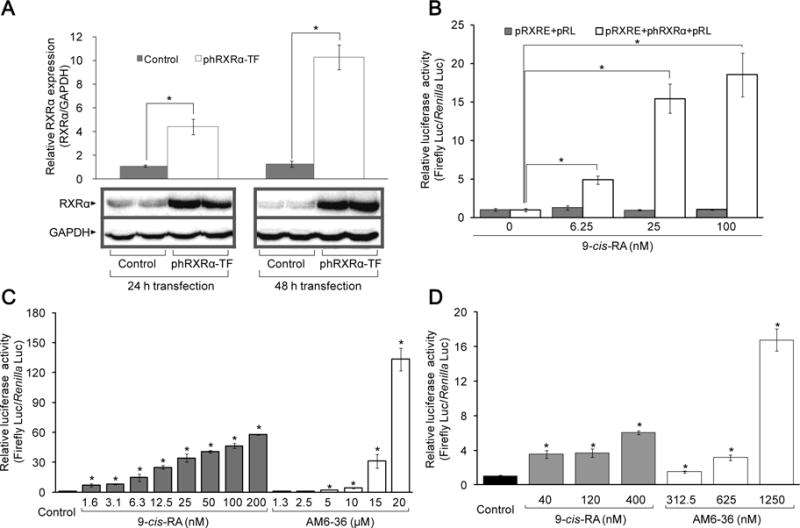
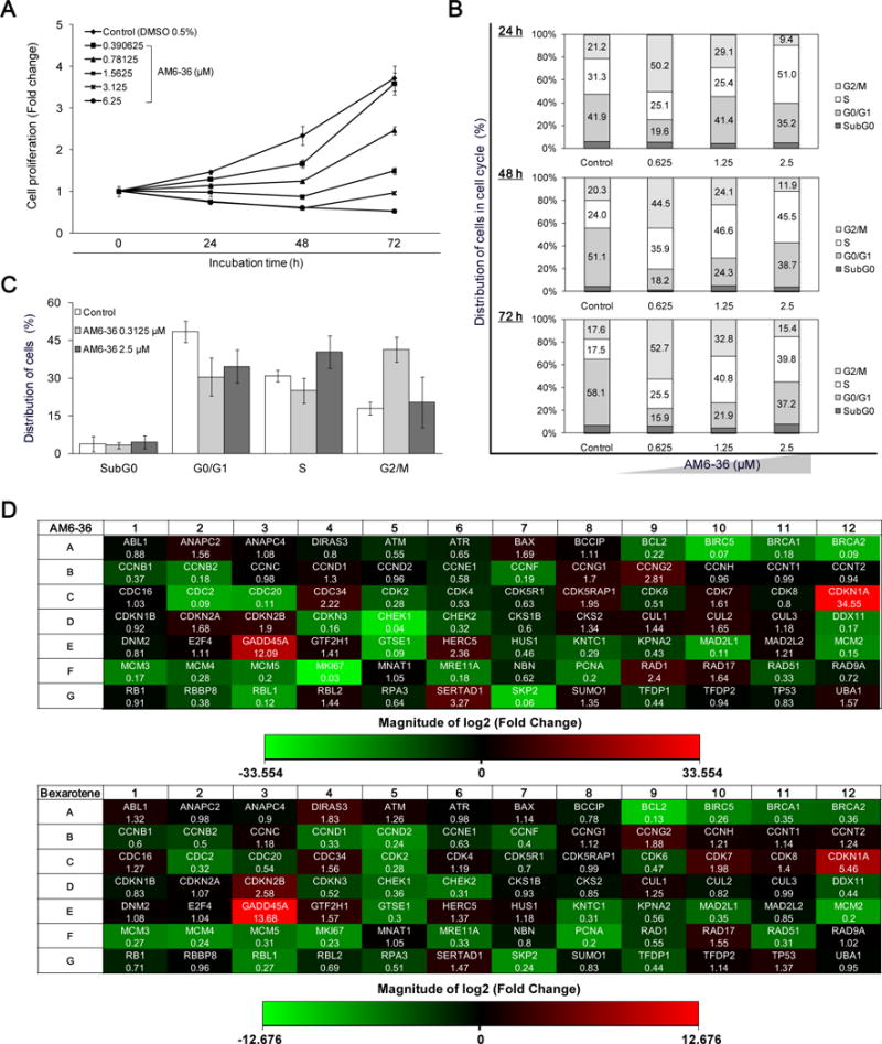
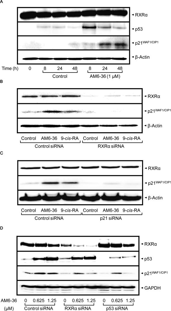
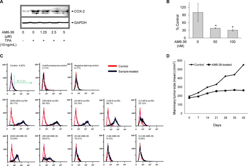
Retinoid X receptor (RXR) has been targeted for the chemoprevention and treatment of cancer. To discover potential agents acting through RXRs, we utilized an RXR response element (RXRE)-luciferase reporter gene assay. Following extensive screening, 3-amino-6-(3-aminopropyl)-5,6-dihydro-5,11-dioxo-11H-indeno[1,2-c]isoquinoline dihydrochloride (AM6-36) was found to induce RXRE-luciferase activities. AM6-36 inhibited COX-2 expression and anchorage-independent growth with 12-O-tetradecanoylphorbol 13-acetate-stimulated JB6 Cl41 cells, induced the expression of CD38 in HL-60 cells, and attenuated the growth of N-methyl-N-nitrosourea-induced mammary tumors in rats. Consistent with other reports describing the antiproliferative effects of RXR agonists in breast cancers, AM6-36 showed growth inhibition with cultured MCF7 breast cancer cells, accompanied by G(2)/M-phase arrest at lower concentrations and enhanced S-phase arrest at higher concentrations. On the basis of DNA microarray analysis, AM6-36 upregulated the expression of CDKN1A, a target gene of RXR, by 35-fold. In accord with this response, the expression of the corresponding protein, p21(WAF1/CIP1), was increased in the presence of AM6-36. Induction of p21 by AM6-36 was abrogated following transient knockdown of RXRα, demonstrating that the effect of AM6-36 on the expression of p21 is closely related to modulation of RXRα transcriptional activity. Intestinal permeability was suggested with Caco-2 cells and limited metabolism resulted when AM6-36 was incubated with human liver microsomes. Oral administration with rats resulted in 0.8 μg/mL, 4.3 μg/g, and 0.3 μg/g in serum, liver, and mammary gland, respectively. In sum, these data suggest that AM6-36 is a promising lead for the treatment or prevention of breast cancer and provide a strong rationale for testing in more advanced antitumor systems.


Figures

Figure 1
Figure 1
Synthesis of AM6-36 and structurally-related indenoisoquinolines.
Figure 2
Figure 2
Effect of 9-cis-RA and AM6-36 on RXR transcriptional activity. A, MCF7 cells were transfected with empty vector (control) or phRXRα (phRXRα-TF) for 24 and 48 h. Total protein lysate (35 μg) of each sample was separated on 12% SDS-PAGE and immuneblotted with anti-RXRα antibody or anti-GAPDH antibody. The mean ± standard deviation (SD) of the band density of RXRα relative to internal control GAPDH of each group (n=3) is shown as a bar graph. Representative Western blot data are presented under the graph. BD, Cells were transiently transfected with plasmids for 24 h, then, further incubated with 9-cis-RA or AM6-36 for 24 h. Cells were lysed and dual luciferase activity was measured. Results were expressed as mean ± SD (n=4). Each mean value was calculated by fold change over control after normalizing ratios of relative light units (RLU) produced by firefly luciferase to RLU produced by Renilla luciferase (Firefly Luc/Renilla Luc). B, COS-1 cells co-transfected with pRXRE (100 ng/well) and pRL (5 ng/well), or pRXRE (100 ng/well), phRXRα (50 ng/well), and pRL (7.5 ng/well) were treated with various concentrations of 9-cis-RA. C, COS-1 cells co-transfected with pRXRE (100 ng/well), phRXRα (50 ng/well) and pRL (7.5 ng/well) were treated with various concentrations of 9-cis-RA or AM6-36. D, MCF7 cells co-transfected with pRXRE (100 ng/well) and pRL (5 ng/well) were incubated with the indicated concentrations of 9-cis-RA or AM6-36. *P value less than 0.05 was considered statistically significant.
Figure 3
Figure 3
Induction of RXRE-luciferase activity and inhibition of cell proliferation accompanied with cell cycle arrest by indenoisoquinolines accompanied by significant changes in cell cycle regulatory genes by AM6-36 treatment in MCF7 breast cancer cells. A, The proliferative state of cells was evaluated with the SRB assay. Cells were incubated for 24 h, 48 h, and 72 h. Results are presented as fold increases of absorbance at 515 nm over control at a starting point (0 h). B and C, MCF7 cells were seeded at a density of 50 × 104 cells in a 10 cm cell culture dish and incubated for 48 h. After incubation with AM6-36 for each time period, 10,000 of cells were analyzed for DNA content and the distribution of cells in each phase was estimated by using ModFit LT™ (Verity Software House, Topsham, ME). D, MCF7 cells were seeded at a density of 50 × 104 cells in a 10 cm cell culture dish and incubated for 48 h. Cells were treated with vehicle (DMSO) or serially diluted AM6-36 for 24 h. RNA was extracted from cells and reverse transcribed to cDNA. Then, cDNA was mixed with RT2 qPCR Master Mix and 25 μl were placed in each well of the array plate. Real-time quantitative PCR was performed using SyBR Green detection system. The thermal cycling condition was at 95°C for 10 min and then 40 cycles at 95°C for 15 sec followed by 60°C for 1 min. Based on the ΔΔCt method, the fold-changes between the control group and sample-treated groups were analyzed using the web-based software provided by SABiosciences (RT2 Profiler PCR Array Data Analysis). Fold changes of 84 genes induced by AM6-36 or bexarotene were determined and presented as heat maps (Red: up-regulation, Green: down-regulation).
Figure 4
Figure 4
Increased protein expression of p53 and p21 by AM6-36, and altered expression of p21 by knock-down of RXRα or p53 in MCF7 cells. A, MCF7 cells were seeded at a density of 10 × 104 cells in 6-well culture plates and incubated for 24 h. Cells were treated with 1 μM AM6-36 for the indicated time points. Cell lysates were prepared and subjected to Western blot analysis. BD, After transient transfection with the indicated siRNAs, MCF7 cells were treated with vehicle (0.1% DMSO), AM6-36 (1 μM, B and C; 0.625, 1, or 1.25 μM, D) or 9-cis-RA (1 μM, B and C) for an additional 24 h. Cell lysates were prepared and subjected to Western blot analysis.
Figure 5
Figure 5
Evaluation of chemopreventive potential of AM6-36. A, JB6 Cl41 cells were stimulated by treatement with TPA (10 ng/ml) in the presence or absence of AM6-36 for 15 h. Whole cell lysates were prepared and COX-2 expression was determined by Western blot analysis. β-Actin was used as an internal control. B, JB6 Cl41 cells were suspended in 50 μl of 0.4% agar complete MEM medium over 100 μl of 0.8% agar complete MEM medium. Cells were exposed to 5 nM TPA with or without AM6-36 at 37°C in a humidified atmosphere with 5% CO2 for 28 days. At the end of the incubation, cell quantification solution was added to each well and further incubated at 37°C for 4 h. Absorbance was measured at 490 nm and the data are shown as % transformation activity relative to TPA-treated control. C, HL-60 cells were incubated with serially diluted 9-cis-RA or AM6-36 (2000, 200, 20, 2, 0.2 nM) for 96 h. Cells were stained with anti-CD38-FITC the intensity of FITC was measured using flow cytometry. FL-1 (+) indicates % of cell population which possess FITC fluorescence, the expression of CD38. D, Effect of AM6-36 (10 mg/kg) on MNU-induced mammary tumors in Sprague-Dawley rats. The plots summarize average tumor size in control (closed triangles) and AM6-36-treated (closed squares) animals (N=10). P=0.1; Mann-Whitney test.
Figure 6
Figure 6
Protein modeling of AM6-36 or 9-cis-RA with RXRα. A, Hypothetical structure of AM6-36 (red) bound to RXRα. B, Overlap of AM6-36 with the crystal structure of 9-cis-RA-RXR complex. AM6-36 is colored red and 9-cis-RA is colored green. The structures are programmed for wall-eyed (relaxed) viewing.

Similar articles

Cited by

References

    1. Mangelsdorf DJ, Ong ES, Dyck JA, Evans RM. Nuclear receptor that identifies a novel retinoic acid response pathway. Nature. 1990;345:224–9. - PubMed
    1. Szanto A, Narkar V, Shen Q, Uray IP, Davies PJ, Nagy L. Retinoid X receptors: X-ploring their (patho)physiological functions. Cell Death Differ. 2004;11:S126–43. - PubMed
    1. Mangelsdorf DJ, Evans RM. The RXR heterodimers and orphan receptors. Cell. 1995;83:841–50. - PubMed
    1. Wolf G. Is 9-cis-retinoic acid the endogenous ligand for the retinoic acid-X receptor? Nutr Rev. 2006;64:532–8. - PubMed
    1. Germain P, Chambon P, Eichele G, et al. International Union of Pharmacology. LXIII. Retinoid X receptors. Pharmacol Rev. 2006;58:760–72. - PubMed

Publication types

MeSH terms

Substances