. 2011 Jun;79(6):2182-92.
doi: 10.1128/IAI.01277-10. Epub 2011 Apr 4.

Discovery of Salmonella virulence factors translocated via outer membrane vesicles to murine macrophages

Affiliations

Discovery of Salmonella virulence factors translocated via outer membrane vesicles to murine macrophages

Hyunjin Yoon et al. Infect Immun. 2011 Jun.

Abstract

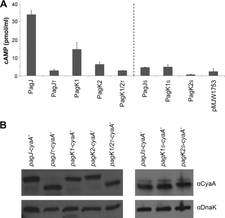
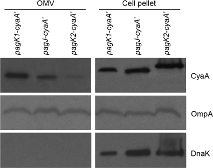
Salmonella enterica serovar Typhimurium, an intracellular pathogen and leading cause of food-borne illness, encodes a plethora of virulence effectors. Salmonella virulence factors are translocated into host cells and manipulate host cellular activities, providing a more hospitable environment for bacterial proliferation. In this study, we report a new set of virulence factors that is translocated into the host cytoplasm via bacterial outer membrane vesicles (OMV). PagK (or PagK1), PagJ, and STM2585A (or PagK2) are small proteins composed of ∼70 amino acids and have high sequence homology to each other (>85% identity). Salmonella lacking all three homologues was attenuated for virulence in a mouse infection model, suggesting at least partial functional redundancy among the homologues. While each homologue was translocated into the macrophage cytoplasm, their translocation was independent of all three Salmonella gene-encoded type III secretion systems (T3SSs)-Salmonella pathogenicity island 1 (SPI-1) T3SS, SPI-2 T3SS, and the flagellar system. Selected methods, including direct microscopy, demonstrated that the PagK-homologous proteins were secreted through OMV, which were enriched with lipopolysaccharide (LPS) and outer membrane proteins. Vesicles produced by intracellular bacteria also contained lysosome-associated membrane protein 1 (LAMP1), suggesting the possibility of OMV convergence with host cellular components during intracellular trafficking. This study identified novel Salmonella virulence factors secreted via OMV and demonstrated that OMV can function as a vehicle to transfer virulence determinants to the cytoplasm of the infected host cell.


Figures

Fig. 1.
Fig. 1.
PagK homologues are translocated independent of Salmonella pathogenicity island 1 (SPI-1) or SPI-2 type III secretion system (T3SS) and the flagellar system. Macrophages were infected with Salmonella strains expressing PagK1, PagK2, or PagJ-CyaA′ in the presence (+) or absence (−) of SPI-1/2 T3SS and flagella for 18 h. (A and B) Translocation of CyaA′ fusions into the macrophage cytosol was measured by the cAMP assay (A), and their expression levels were compared in parallel by Western blotting using anti-CyaA antibody (αCyaA) (B). DnaK levels were used to normalize protein levels. In order to confirm that SPI-1 and SPI-2 secretion systems were disrupted by deleting invA and ssaK, SipA (SPI-1 effector) and SseJ (SPI-2 effector) were tagged with CyaA′, and their translocation (A) and expression (B) were examined in the absence of InvA and SsaK, respectively. Note that translocation and expression of SipA-CyaA′ was measured at 1 h postinfection, and the analysis of the other CyaA′-tagged proteins was performed at 18 h postinfection. Salmonella not expressing CyaA′ (strain 14028s) or expressing LacZ-CyaA′ from pMJW1791 (30) did not exhibit an increased cAMP level in panel A as expected. Intriguingly, the level of expression of each PagK homologue increased in the triple mutant background as can be observed.
Fig. 2.
Fig. 2.
N-terminal deletion of PagK homologues abolishes their translocation into the host cells. The full-length PagK homologues and their truncated forms without their predicted N-terminal signal peptides or with their N-terminal signal peptides only were fused to CyaA′ as described in Materials and Methods. Salmonella strains expressing the full-length PagK homologues (PagJ, PagK1, and PagK2) and the truncated derivatives (PagJT and PagK1/PagK2T lacking N-terminal signal peptides; PagJS, PagK1S, and PagK2S harboring only N-terminal signal peptides) were inoculated into RAW264.7 macrophage-like cells. (A and B) Infected macrophage cells were lysed at 6 h postinfection for cAMP assay (A) and Western blot analysis (B). (A)The translocation levels of PagK homologues and their derivatives were compared by measuring intracellular cAMP levels. (B) In parallel, their expression levels were determined by anti-CyaA antibody using the same macrophage lysates used in the cAMP assay. DnaK levels were measured to normalize protein levels loaded in the different lanes. Note that the sequences of PagK1 and PagK2 are identical if the signal sequences have been deleted; therefore, only a single construct, PagK1/2T, is shown for the N-terminally truncated derivatives of PagK1 and PagK2. Salmonella harboring pMJW1753, which was used as a backbone construct in all CyaA′ fusions in this experiment, was used in the cAMP assay as a negative control.
Fig. 3.
Fig. 3.
PagK2 is colocalized with LPS but not with Salmonella inside macrophages. Macrophages were infected with Salmonella expressing PagK2-CyaA′ for 18 h. (A to D) The fixed cells were incubated with rabbit anti-CyaA antibody (B) and mouse anti-LPS antibody (A) overnight and treated with Alexa Fluor 647-conjugated anti-rabbit IgG (B), Alexa Fluor 350-conjugated anti-mouse IgG (A), and FITC-conjugated cholera toxin B subunit (CTB) (D). Salmonella expresses the red fluorescent protein “Tomato” from pWKS30-Tomato plasmid (C). (A) LPS staining in the blue channel; (B) CyaA′ staining in the yellow channel; (C) Salmonella with fluorescence from the the red fluorescent protein Tomato; (D) plasma membrane staining in the green channel using CTB; (E) merged image of LPS, CyaA′, and Salmonella; (F) merged image of LPS, CyaA′, CTB, and Salmonella. The sites of colocalization of PagK2 with LPS are indicated by white arrows in panel F. Note that a bleb-like protuberance from the host cell (indicated by the white arrow in panel D) encloses PagK2 and LPS (as shown in panel F), while the bacteria are present in the spacious vacuole seen as a large red vacuole at the top of the cell as depicted.
Fig. 4.
Fig. 4.
PagK homologues are present in isolated outer membrane vesicles (OMV). Salmonella strains expressing CyaA′-tagged PagK1, PagJ, and PagK2 (C-terminal translational fusions) were cultivated on AMM1 medium for 5 h, and vesicles were isolated as described in Materials and Methods. Vesicles produced from 80 ml of bacterial culture (∼24 μg/80 ml) were precipitated with TCA to concentrate proteins. (Left) The vesicle-containing precipitate produced from the initial 80-ml culture supernatant was resuspended in 1× Laemmli sample buffer and loaded in a lane of the SDS-polyacrylamide gel (OMV fraction). (Right) Bacterial cells (5 × 10⁷) were pelleted and loaded onto the SDS-polyacrylamide gel (cell pellet fraction). PagK1, PagJ, and PagK2 were immunoblotted using anti-CyaA antibody. OmpA, a highly expressed outer membrane protein, was used as a control for the presence of outer membrane vesicles. DnaK, a cytosolic protein, was used to verify that no bacterial lysis occurred during the OMV isolation procedure. Note that the molecular weight of PagK2 is approximately the same as PagK1 and PagJ in OMV fraction but not within whole bacterial cells (compare the molecular weight of PagK2 with those of PagK1 and PagJ in the OMV and cell pellet fractions). We speculate that signal sequences present in these effectors may contribute to the difference in molecular weights between the left- and right-hand panels.
Fig. 5.
Fig. 5.
Localization of PagK homologues in intact unsectioned OMV. Vesicles were isolated from the culture of Salmonella pagK2-cyaA′ strain grown on AMM1 medium as described in Materials and Methods. Isolated vesicles were fixed and loaded onto Formvar/carbon-coated grids without sectioning vesicles. (A and B) The grids were incubated with a solution containing rabbit anti-CyaA antibody (A) or rabbit anti-LPS antibody (B) overnight and then treated with 15-nm-gold-conjugated anti-rabbit IgG. The micrographs are representative of the vesicles examined. (A) PagK2-CyaA′; (B) LPS. Vesicles isolated from cultures of pagJ-cyaA′ and pagK1-cyaA′ strains also showed similar distribution patterns of anti-CyaA and anti-LPS antibodies.
Fig. 6.
Fig. 6.
Immunogold electron microscopy to locate PagK homologues inside macrophages. Macrophages were infected with Salmonella CyaA′ fusion strains for 10 h and fixed with 4% paraformaldehyde for transmission electron microscopy. The methods are described in detail in Materials and Methods. CyaA′-tagged PagK homologues were detected using rabbit anti-CyaA antibody and 15-nm-gold-conjugated anti-rabbit IgG (small black beads indicated by small black arrows). LPS was labeled with 25-nm-gold-conjugated anti-LPS antibody (large black beads). Salmonella and OMV are indicated by S and V, respectively. (A to F) The micrographs show macrophages infected with Salmonella strain pagJ-cyaA′ (A and B), pagK1-cyaA′ (C and D), and pagK2-cyaA′ (E and F).
Fig. 7.
Fig. 7.
Modification of OMV during translocation into the cytoplasm. (A and B) Uninfected macrophages (A) and Salmonella-infected macrophages (B) (10 h after infection) were examined after ultrathin sectioning. The grids were treated with 15-nm-gold-conjugated anti-LAMP1 antibody and 25-nm-gold-conjugated anti-LPS antibody as described in Materials and Methods. Based on the examination of 100 cells from the thin sections prepared in parallel, LAMP1 molecules (small black beads indicated by black arrows) were more plentiful in Salmonella-infected macrophages and appeared to be enriched in OMV released from the Salmonella-containing vacuole. Salmonella and OMV are labeled with S and V, respectively. (A) RAW264.7 macrophage-like cells; (B) RAW264.7 cells infected with S. Typhimurium 14028s.

Similar articles

Cited by

References

    1. Amano A., Takeuchi H., Furuta N. 2010. Outer membrane vesicles function as offensive weapons in host-parasite interactions. Microbes Infect. 12:791–798 - PubMed
    1. Asensio C. J., et al. 2011. Outer membrane vesicles obtained from Bordetella pertussis Tohama expressing the lipid A deacylase PagL as a novel acellular vaccine candidate. Vaccine 29:1649–1656 - PubMed
    1. Ayala B. P., et al. 2001. The pilus-induced Ca2+ flux triggers lysosome exocytosis and increases the amount of Lamp1 accessible to Neisseria IgA1 protease. Cell. Microbiol. 3:265–275 - PubMed
    1. Bakke H., et al. 2001. Meningococcal outer membrane vesicle vaccine given intranasally can induce immunological memory and booster responses without evidence of tolerance. Infect. Immun. 69:5010–5015 - PMC - PubMed
    1. Beatty W. L., et al. 2000. Trafficking and release of mycobacterial lipids from infected macrophages. Traffic 1:235–247 - PubMed

Publication types

Substances

LinkOut - more resources