. 2011 Apr 19;108(16):6503-8.
doi: 10.1073/pnas.1015449108. Epub 2011 Apr 4.

Dual origin of mesenchymal stem cells contributing to organ growth and repair

Affiliations

Dual origin of mesenchymal stem cells contributing to organ growth and repair

Jifan Feng et al. Proc Natl Acad Sci U S A. .

Abstract

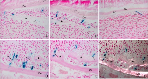
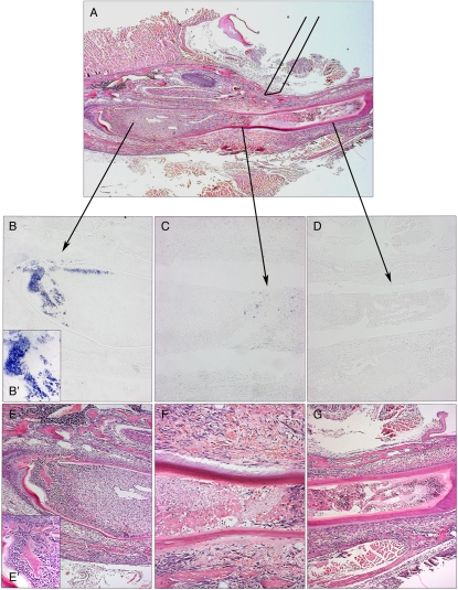
In many adult tissues, mesenchymal stem cells (MSCs) are closely associated with perivascular niches and coexpress many markers in common with pericytes. The ability of pericytes to act as MSCs, however, remains controversial. By using genetic lineage tracing, we show that some pericytes differentiate into specialized tooth mesenchyme-derived cells--odontoblasts--during tooth growth and in response to damage in vivo. As the pericyte-derived mesenchymal cell contribution to odontoblast differentiation does not account for all cell differentiation, we identify an additional source of cells with MSC-like properties that are stimulated to migrate toward areas of tissue damage and differentiate into odontoblasts. Thus, although pericytes are capable of acting as a source of MSCs and differentiating into cells of mesenchymal origin, they do so alongside other MSCs of a nonpericyte origin. This study identifies a dual origin of MSCs in a single tissue and suggests that the pericyte contribution to MSC-derived mesenchymal cells in any given tissue is variable and possibly dependent on the extent of the vascularity.


Conflict of interest statement

The authors declare no conflict of interest.

Figures

Fig. 1.
Fig. 1.
Pericytes can differentiate into odontoblasts following damage. NG2creER;Rosa26R pups were given one intraperitoneal injection of tamoxifen at P2 (4 mg/30 g body weight), and at 54 h after tamoxifen injection, mandibles on the left side were damaged by using a needle and the pups were culled 2 d (A and B) and 4 d (D and E) later. Right sides were used as controls (no damage) (C). After 2 d, the site of damage showed an eosinophilic cell matrix compatible with immature predentin (marked with asterisk) with close contact with NG2 LacZ-positive (NG2-positive) elongated odontoblast-like cells (A and B). At 4 d after damage, the number of LacZ-positive odontoblasts in close contact with the lesion greatly increased, showing clear signs of further differentiation with elongated bodies and long processes inside the reparative dentine (asterisk) canaliculus (D and E). Polarized light microscopy showed collagen fibrils in the reparative areas (asterisk), parallel to the odontoblast longitudinal axis (F). Black arrows show examples of LacZ-positive odontoblasts extending stubby processes inside the dentin matrix. De, dentin; PD, predentin.
Fig. 2.
Fig. 2.
Pericytes can respond to injury locally, are involved in tooth development, and are slow-cycling cells. By culturing the X-LacZ4 incisors following damage, and thus eliminating the circulatory system, an increase in the number of LacZ-positive pericytes was evident in both pulp body (A) and cervical loop parts (B), compared with the control incisors (A′ and B′). These cells were characteristically dispersed throughout the pulp tissue, losing their organization within the main body plexus in the pulp body (A and A′), and were intensely concentrated on the bottom edge of the cervical loop area (B and B′), where a vessel-rich area is present. Coexpression of proliferation marker PH3 and LacZ-positive (NG2-positive) pericytes following in vivo damage (C and C′) showed that proliferating pericytes (black arrows) are located on blood vessels extending toward the damaged area (C′). During tooth development (bud to bell stages) in X-LacZ4 mice, LacZ-positive cells are found under the condensing mesenchyme (D) and in the blood vessels close to the forming tooth (E). When colocation of migrating slow-cycling cells and pericytes were analyzed by transplantation experiments associated with nucleoside (i.e., IdU) administration, some cells that migrated from the X-LacZ4 host were both LacZ-positive (pericytes, black stained cells) and IdU positive (slow-cycling, red stained cells) (G and H). These cells were found in the blood vessel-rich cervical loop area of incisors (F and G). Consecutive sections showed that these cells were rare but clustered (G), suggesting a spatial organization. High magnification showed colocalization of IdU and LacZ-positive pericytes under fluorescence (H) and bright field (I).
Fig. 3.
Fig. 3.
Directed cell migration toward tissue damage visualized by DiI labeling of incisor mesenchyme pulp cells. Cells labelled in cervical loop area (AC), damaged pulp cells area (AC), damaged pulp body (DF), and control teeth with no damage (GI). Arrows indicate position of tooth damage. A lesion separating the cervical loop and the pulp body was provoked in incisors, and, 48 h later, mesenchymal cells were capable of migrating toward the site of the damage (AC) whereas the mesenchymal cells from the pulp body showed no migration (DF). In the absence of the lesion, no directed migration of cervical loop mesenchymal cells was observed (GI).
Fig. 4.
Fig. 4.
Differentiation into Dspp-positive preodontoblasts following damage. Following damage to three separate sites, Dspp expression was visualized by in situ hybridization to reveal early odontoblast differentiation. Damage directly to the cervical loop area showed extensive expression of Dspp after 2 d (B and B′), whereas only a few expressing cells were visible in the damaged region midway along the length of the tooth (C). At the tip of the tooth, no Dspp expression was evident (D). Note that evidence of an inflammatory response can be seen in the tip (G), body (F), and cervical loop cells (E), but presence of osteodentin is evident in only the cervical loop (E′).

References

    1. Friedenstein AJ, Chailakhyan RK, Latsinik NV, Panasyuk AF, Keiliss-Borok IV. Stromal cells responsible for transferring the microenvironment of the hemopoietic tissues. Cloning in vitro and retransplantation in vivo. Transplantation. 1974;17:331–340. - PubMed
    1. Caplan AI. Mesenchymal stem cells. J Orthop Res. 1991;9:641–650. - PubMed
    1. Pittenger MF, et al. Multilineage potential of adult human mesenchymal stem cells. Science. 1999;284:143–147. - PubMed
    1. Zuk PA, et al. Human adipose tissue is a source of multipotent stem cells. Mol Biol Cell. 2002;13:4279–4295. - PMC - PubMed
    1. Toma JG, et al. Isolation of multipotent adult stem cells from the dermis of mammalian skin. Nat Cell Biol. 2001;3:778–784. - PubMed

Publication types