. 2011 Apr 19;108(16):6486-91.
doi: 10.1073/pnas.1019062108. Epub 2011 Apr 4.

Deactivation of Akt by a small molecule inhibitor targeting pleckstrin homology domain and facilitating Akt ubiquitination

Affiliations

Deactivation of Akt by a small molecule inhibitor targeting pleckstrin homology domain and facilitating Akt ubiquitination

Hakryul Jo et al. Proc Natl Acad Sci U S A. .

Abstract

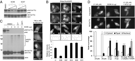
The phosphatidylinositol-3,4,5-triphosphate (PIP3) binding function of pleckstrin homology (PH) domain is essential for the activation of oncogenic Akt/PKB kinase. Following the PIP3-mediated activation at the membrane, the activated Akt is subjected to other regulatory events, including ubiquitination-mediated deactivation. Here, by identifying and characterizing an allosteric inhibitor, SC66, we show that the facilitated ubiquitination effectively terminates Akt signaling. Mechanistically, SC66 manifests a dual inhibitory activity that directly interferes with the PH domain binding to PIP3 and facilitates Akt ubiquitination. A known PH domain-dependent allosteric inhibitor, which stabilizes Akt, prevents the SC66-induced Akt ubiquitination. A cancer-relevant Akt1 (e17k) mutant is unstable, making it intrinsically sensitive to functional inhibition by SC66 in cellular contexts in which the PI3K inhibition has little inhibitory effect. As a result of its dual inhibitory activity, SC66 manifests a more effective growth suppression of transformed cells that contain a high level of Akt signaling, compared with other inhibitors of PIP3/Akt pathway. Finally, we show the anticancer activity of SC66 by using a soft agar assay as well as a mouse xenograft tumor model. In conclusion, in this study, we not only identify a dual-function Akt inhibitor, but also demonstrate that Akt ubiquitination could be chemically exploited to effectively facilitate its deactivation, thus identifying an avenue for pharmacological intervention in Akt signaling.


Conflict of interest statement

The authors declare no conflict of interest.

Figures

Fig. 1.
Fig. 1.
Cell-based screening identifies a compound that directly facilitates Akt ubiquitination. (A) HeLa-PH-EGFP cells were treated with SC66 (4 μg/mL) for 1 h and were stained with the γ-tubulin antibody (red). Arrows indicate the colocalization of PH-EGFP with γ-tubulin (Left). HEK293 cells transfected with the indicated EGFP fusion proteins were treated with SC66 alone or together with IGF1 (20 ng/mL) or wortmannin (200 nM). After 1 h, pericentrosomal localization was visualized in live cells. Arrows indicate the pericentrosomal region (Middle). HEK293 cells stably expressing PH-EGFP were transfected with the C-terminal V5/His tagged Akt1. Following treatment with SC66 for 1 h, the fixed cells were stained for Akt1. Colocalization with PH-EGFP in pericentrosomal region is indicated by arrows (Right). (B) HEK293T cells grown in serum-rich medium were treated with different amounts of SC66 for 1 h. The whole-cell lysates were analyzed with the indicated antibodies. (C) HEK293-Akt1 cells were treated with SC66 (4 μg/mL), MG132 (10 μg/mL), or a combination of the two for 4 h, and the whole-cell lysates were analyzed by a monoclonal V5 antibody (Upper). The cell lysates from SC66-treated cells were supplemented with 50 mM imidazole followed by incubation with Ni-NTA beads in the presence or absence of 1 mM DTT. The bead-bound fraction was resolved on a SDS/PAGE and blotted with the V5 antibody for Akt1. The same membrane was autoclaved and sequentially blotted with a rabbit polyclonal antibody against ubiquitin (Lower). (D) HEK293 cell lysates were subjected to in vitro ubiquitination assay with or without SC66 for different times (Left). The Akt1 immune complex was treated with DMSO or SC66 for 1 h, and washed three times with the buffer. The resulting immune complex was subjected to in vitro ubiquitination with fresh HEK cell lysates (Right). (E) Inhibitory effect of an allosteric Akt inhibitor, AKTi-VIII, on the SC66-induced in vitro ubiquitination of Akt1.
Fig. 2.
Fig. 2.
SC66 functionally inhibits a cancer-relevant Akt1 (e17k) mutant. (A) HEK293 cells stably expressing Akt1 (e17k) mutant were treated with different amounts (2, 4, 8 μg/mL) of SC66 or SC67 for 1 h, followed by Western blot for phospho-Akt (S473). (B) HeLa cells expressing PH (e17k)–EGFP were treated with LY294002 (40 μM) or indicated compounds (4 μg/mL) in the presence of IGF1. The relative intensity of membrane PH (e17k)–EGFP between time 0 and 60 min was quantified. The arrows indicate the pericentrosomal accumulation of PH(e17k)–EGFP. (C) HEK293-Akt1 (e17k) cells were pretreated with LY294002 or AKTi-VIII for 30 min followed by SC66 treatment for an additional 2 h, and the cytosolic extracts were analyzed for phospho-Akt and ubiquitinated Akt1 (e17k) (Left). HeLa cells transfected with Akt1 (e17k) were treated with AKTi-VIII, SC66, or a combination of the two for 2 h. The representative immunostaining for Akt1 (e17k) is shown (Right). (D) HeLa cells cotransfected with EGFP-Foxo and Akt1 (e17k) were treated with different amounts of LY294002, SC66, or a combination of the two. The coexpressing cells were identified by immunostaining for Akt1 (e17k). The relative intensity of cytosolic versus nuclear EGFP-Foxo was determined and scored as cytosolic (>1.1), equal (1.1–0.9), or nuclear (<0.9; Right).
Fig. 3.
Fig. 3.
SC66 enhances cancer cell death mediated by PI3K inhibition. (A) Confluent HeLa cells were scratched and, following a 10-min recovery, the indicated chemicals were added and incubated for 6 h. The quantification is presented in Fig. S11A. (B) Analysis of time-lapse live cell imaging of HeLa cells undergoing mitosis or apoptosis in the presence of indicated chemicals. HeLa cells treated with each chemical were imaged every 15 min for the duration of 14 h. Each frame was sequentially analyzed to identify cells entering mitosis (dotted circle) or undergoing apoptosis (dotted rectangle) within this time period. The percentage of these cell numbers in reference to initial cell numbers in each condition was presented. (C) HeLa cells transfected with EGFP-Foxo were treated with different amounts of LY294002 for 1 h (Top) or incubated for 16 h followed by an additional 1-h incubation with LY294002 or SC66. The intensity of cytosolic and nuclear EGFP-Foxo was determined, and the percentage of cells with nuclear EGFP-Foxo was presented (*P < 0.05, Student t test). (D) Representative pictures of time-lapse (14 h) live cell imaging of HeLa cells treated with LY294002, SC66, or a combination of the two. The quantification is presented in Fig. S13C. FACS analysis of HeLa cells treated with LY294002, SC66, or a combination of the two for 20 h. The quantification is presented in Fig. S13D.
Fig. 4.
Fig. 4.
SC66 manifests anticancer activity in vitro and in vivo. (A) HEK293T cells grown in serum-rich medium were treated with the indicated amounts of each compound for 1 h. The phosphorylation levels of Akt and its target proteins were examined. (B) HEK293T cells were treated with the indicated amounts of each compound for 16 h, and cell viability was determined by MTT assay. (C) Inhibitory effect of SC66 toward colony formation of HEK293T cells on soft agar. Representative image of wells following a 3-week culture in the presence of different amounts of SC66 is shown. Quantification is from three independent experiments. (D) HEK293T cells were inoculated into the nude mice, and the mice were treated with vehicle alone or two different concentrations of SC66. The growth of tumors was measured at the indicated time points. Representative images of dissected tumors after 28 d are shown. P values between paired groups (Student t test) are as follows: control vs. SC66 15 mg/kg, P = 0.0209; control vs. SC66 30 mg/kg, P = 0.0190; and SC66 15 mg/kg vs. SC66 30 mg/kg, P = 0.0121.

References

    1. Bunney TD, Katan M. Phosphoinositide signalling in cancer: Beyond PI3K and PTEN. Nat Rev Cancer. 2010;10:342–352. - PubMed
    1. Engelman JA, Luo J, Cantley LC. The evolution of phosphatidylinositol 3-kinases as regulators of growth and metabolism. Nat Rev Genet. 2006;7:606–619. - PubMed
    1. Guertin DA, Sabatini DM. Defining the role of mTOR in cancer. Cancer Cell. 2007;12:9–22. - PubMed
    1. Bozulic L, Hemmings BA. PIKKing on PKB: Regulation of PKB activity by phosphorylation. Curr Opin Cell Biol. 2009;21:256–261. - PubMed
    1. Engelman JA. Targeting PI3K signalling in cancer: Opportunities, challenges and limitations. Nat Rev Cancer. 2009;9:550–562. - PubMed

Publication types

MeSH terms