. 2011 May;60(5):1483-92.
doi: 10.2337/db10-0869. Epub 2011 Apr 4.

Ablation of PI3K p110-α prevents high-fat diet-induced liver steatosis

Affiliations

Ablation of PI3K p110-α prevents high-fat diet-induced liver steatosis

Mohar Chattopadhyay et al. Diabetes. 2011 May.

Abstract

Objective: To determine whether the phosphoinositide 3-kinase (PI3K) catalytic subunits p110-α and p110-β play a role in liver steatosis induced by a high-fat diet (HFD).

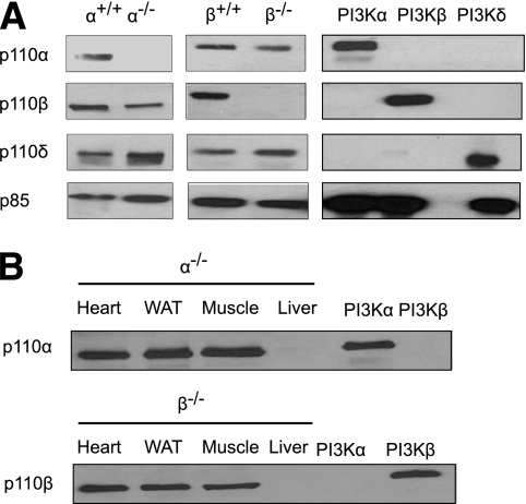
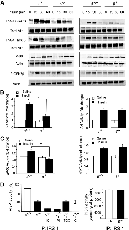
Research design and methods: Liver-specific p110-α and p110-β knockout mice and control animals for each group were fed an HFD or normal chow for 8 weeks. Biochemical assays and quantitative real-time PCR were used to measure triglyceride, expression of lipogenic and gluconeogenic genes, and activity of protein kinases downstream of PI3K in liver lysates. Fatty acid uptake and incorporation into triglycerides were assessed in isolated hepatocytes.

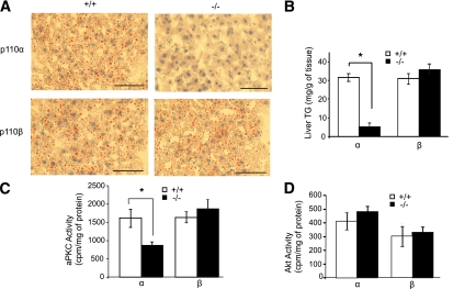
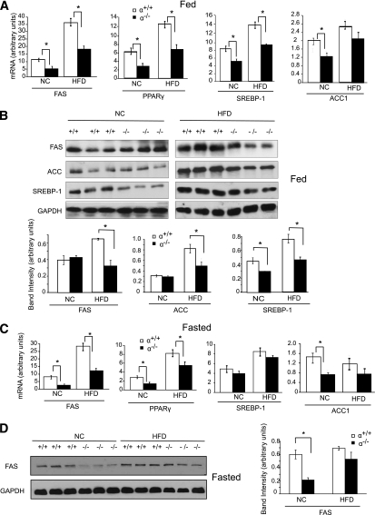
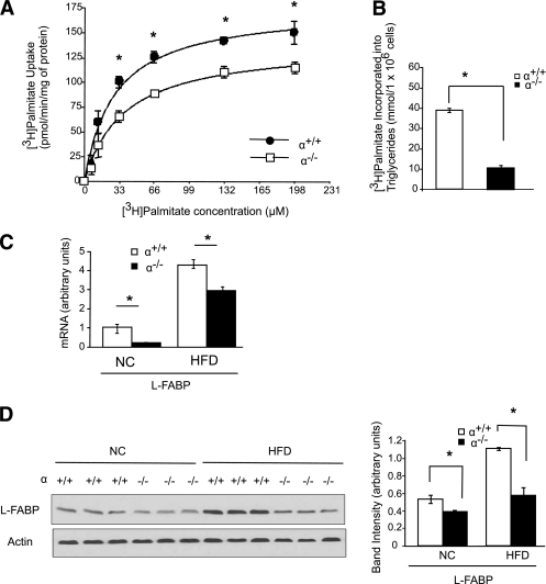
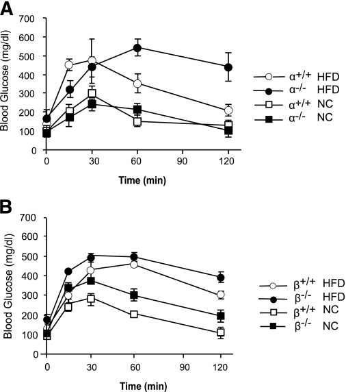
Results: Hepatic triglyceride levels in HFD-fed p110-α(-/-) mice were 84 ± 3% lower than in p110-α(+/+) mice, whereas the loss of p110-β did not significantly alter liver lipid accumulation. p110-α(-/-) livers also showed a reduction in atypical protein kinase C activity and decreased mRNA and protein expression of several lipogenic genes. Hepatocytes isolated from p110-α(-/-) mice exhibited decreased palmitate uptake and reduced fatty acid incorporation into triglycerides as compared with p110-α(+/+) cells, and hepatic expression of liver fatty acid binding protein was lower in p110-α(-/-) mice fed the HFD as compared with controls. Ablation of neither p110-α nor p110-β ameliorated glucose intolerance induced by the HFD, and genes involved in gluconeogenesis were upregulated in the liver of both knockout animals.

Conclusions: PI3K p110-α, and not p110-β, promotes liver steatosis in mice fed an HFD. p110-α might exert this effect in part through activation of atypical protein kinase C, upregulation of lipogenesis, and increased uptake of fatty acids.


Figures

FIG. 1.
FIG. 1.
Ablation of p110-α− and p110-β−PI3K in the liver. A: Class IA PI3Ks in liver lysates from mice of the indicated genotypes were pulled down with a phosphotyrosine peptide and the bound proteins were examined on Western blots probed with the indicated antibodies. Recombinant PI3K-α (p110-α/p85-α), PI3K-β (p110-β/p85-α), and PI3K-δ (p110-δ/p85-α) were loaded as controls. B: Class IA PI3Ks in lysates from hearts, white adipose tissue (WAT), skeletal muscle, and livers of p110-α−/− and p110-β−/− mice were pulled down with a phosphotyrosine peptide, and the bound proteins were examined on Western blots probed with the indicated antibodies. Recombinant PI3K-α and PI3K-β were loaded as controls.
FIG. 2.
FIG. 2.
Insulin signaling in p110-null livers. A: Liver lysates from fasted mice treated intraperitoneally without or with insulin (2 units/kg body wt) for the indicated times were analyzed by Western blotting. B and C: Fasted mice were injected intraperitoneally with saline or insulin (2 units/kg body wt). Livers were collected 15 min later, and lysates were prepared and assayed for Akt (B) or aPKC (C) kinase activity. *P < 0.05 by t test (N = 3 for all groups). D: Mice were fasted overnight and then injected through the inferior vena cava with insulin (2 units/kg body wt). Livers were collected 5 min later, and lysates were subjected to immunoprecipitation with IRS-1 antibody. The immunoprecipitates for p110-α+/+ and p110-α−/− (left panel) were assayed for PI3K activity in the presence of vehicle, 500 nmol/L PI-103 (PI), 50 nmol/L TGX-221 (TGX), or 5 μmol/L IC87114 (IC). Values are normalized to the average PI3K activity in p110-α+/+ samples assayed in the absence of inhibitors. The immunoprecipitates for p110-β+/+ and p110-β−/− (right panel) were assayed without any additions (N = 3 for all groups). Values shown are mean ± SEM.
FIG. 3.
FIG. 3.
Loss of p110-α blocks hepatic steatosis induced by an HFD. Mice were fed a 45 kcal% fat diet for 8 weeks. A: Frozen liver sections (8 µm) were stained with Oil Red O to visualize neutral lipids and counterstained with hematoxylin. Scale bars = 200 µm. B: Liver triglyceride (TG) content. *P < 0.005 by t test (N = 6). C: aPKC kinase activity assayed in liver lysates from mice fasted for 6 h. *P < 0.01 by t test (N = 3). D: Akt kinase activity assayed in liver lysates from mice fasted for 6 h (N = 3). Values are mean ± SEM. (A high-quality digital representation of this figure is available in the online issue.)
FIG. 4.
FIG. 4.
Loss of p110-α affects the expression of genes and proteins regulating lipid metabolism. A: Quantitative RT-PCR analysis of mRNA levels in livers of mice fed ad libitum an HFD or normal chow (NC). *P < 0.05 by t test (N = 6). B: Liver lysates from ad libitum-fed mice were analyzed by Western blotting with the indicated antibodies. Bands were quantified using densitometry. *P < 0.01 by t test. C: Quantitative RT-PCR analysis of mRNA levels in livers of mice fasted for 6 h. *P < 0.05 by t test (N = 6). D: Liver lysates from mice fasted for 6 h were analyzed on Western blots, and the bands were quantified by densitometry. *P < 0.01 by t test. Values are means ± SEM.
FIG. 5.
FIG. 5.
Loss of p110-α attenuates fatty acid uptake in hepatocytes. Hepatocytes were isolated from p110-α+/+ and p110-α−/− mice fed normal chow as described in research design and methods. A: Cells were incubated with increasing amounts of [3H]palmitate/BSA at a 3:1 molar ratio for 1 min. [3H]palmitate uptake into the cell was then measured. *P < 0.001 by t test. B: Hepatocytes were incubated with [3H]palmitate/BSA for 2 h, and [3H]palmitate incorporated into cellular triglycerides was then measured. *P < 0.001 by t test. C: Quantitative RT-PCR analysis of mRNA levels in livers of p110-α+/+ and p110-α−/− mice fed an HFD or normal chow (NC). *P < 0.05 by t test (N = 6). D: Liver lysates were analyzed by Western blotting with the indicated antibodies. Bands were quantified using densitometry. *P < 0.005 by t test. Values are means ± SEM of three independent experiments done in triplicate.
FIG. 6.
FIG. 6.
Glucose intolerance in mice fed an HFD. A and B: Glucose tolerance tests in mice of the indicated genotypes fed an HFD or normal chow (NC) (N = 6 in each group).

References

    1. Angulo P. Nonalcoholic fatty liver disease. N Engl J Med 2002;346:1221–1231 - PubMed
    1. Angulo P. GI epidemiology: nonalcoholic fatty liver disease. Aliment Pharmacol Ther 2007;25:883–889 - PubMed
    1. Inoue M, Ohtake T, Motomura W, et al. Increased expression of PPARgamma in high fat diet-induced liver steatosis in mice. Biochem Biophys Res Commun 2005;336:215–222 - PubMed
    1. Lin J, Yang R, Tarr PT, et al. Hyperlipidemic effects of dietary saturated fats mediated through PGC-1beta coactivation of SREBP. Cell 2005;120:261–273 - PubMed
    1. Oosterveer MH, van Dijk TH, Tietge UJ, et al. High fat feeding induces hepatic fatty acid elongation in mice. PLoS ONE 2009;4:e6066. - PMC - PubMed

Publication types

MeSH terms