

Role of the cytoskeleton in formation and maintenance of angiogenic sprouts
- PMID: 21464572
- PMCID: PMC3080587
- DOI: 10.1159/000324751
Role of the cytoskeleton in formation and maintenance of angiogenic sprouts
Abstract
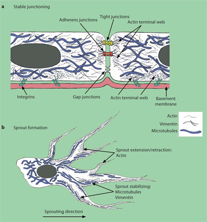
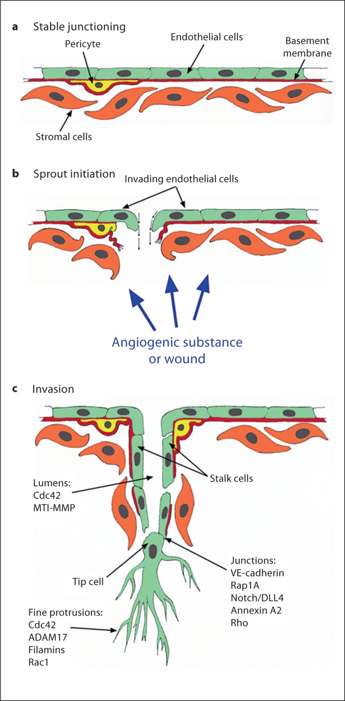
Angiogenesis is the formation of new blood vessels from pre-existing structures, and is a key step in tissue and organ development, wound healing and pathological events. Changes in cell shape orchestrated by the cytoskeleton are integral to accomplishing the various steps of angiogenesis, and an intact cytoskeleton is also critical for maintaining newly formed structures. This review focuses on how the 3 main cytoskeletal elements--microfilaments, microtubules, and intermediate filaments--regulate the formation and maintenance of angiogenic sprouts. Multiple classes of compounds target microtubules and microfilaments, revealing much about the role of actin and tubulin and their associated molecules in angiogenic sprout formation and maintenance. In contrast, intermediate filaments are much less studied, yet intriguing evidence suggests a vital, but unresolved, role in angiogenic sprouting. This review discusses evidence for regulatory molecules and pharmacological compounds that affect actin, microtubule and intermediate filament dynamics to alter various steps of angiogenesis, including endothelial sprout formation and maintenance.
Copyright © 2011 S. Karger AG, Basel.
Figures
References
-
- Carmeliet P. Angiogenesis in health and disease. Nat Med. 2003;9:653–660. - PubMed
-
- Carmeliet P. Manipulating angiogenesis in medicine. J Intern Med. 2004;255:538–561. - PubMed
-
- Ferrara N, Carver-Moore K, Chen H, Dowd M, Lu L, O'Shea KS, Powell-Braxton L, Hillan KJ, Moore MW. Heterozygous embryonic lethality induced by targeted inactivation of the VEGF gene. Nature. 1996;380:439–442. - PubMed
-
- Fong GH, Rossant J, Gertsenstein M, Breitman ML. Role of the Flt-1 receptor tyrosine kinase in regulating the assembly of vascular endothelium. Nature. 1995;376:66–70. - PubMed
-
- Shalaby F, Rossant J, Yamaguchi TP, Gertsenstein M, Wu XF, Breitman ML, Schuh AC. Failure of blood-island formation and vasculogenesis in Flk-1-deficient mice. Nature. 1995;376:62–66. - PubMed
