. 2011;48(5):386-96.
doi: 10.1159/000323474. Epub 2011 Apr 4.

Constitutive expression and enzymatic cleavage of ICAM-1 in the spontaneously hypertensive rat

Affiliations

Constitutive expression and enzymatic cleavage of ICAM-1 in the spontaneously hypertensive rat

Sheng Tong et al. J Vasc Res. 2011.

Abstract

Background/aims: Leukocyte adhesion to the endothelium is abnormal in hypertension. We have recently shown that spontaneously hypertensive rats (SHRs) have circulating leukocytes with enhanced CD18 receptor cleavage. In the current study, we investigate expression levels of its counter receptor, intercellular adhesion molecule (ICAM-1), and its possible proteolytic cleavage in the SHR and control Wistar rat.

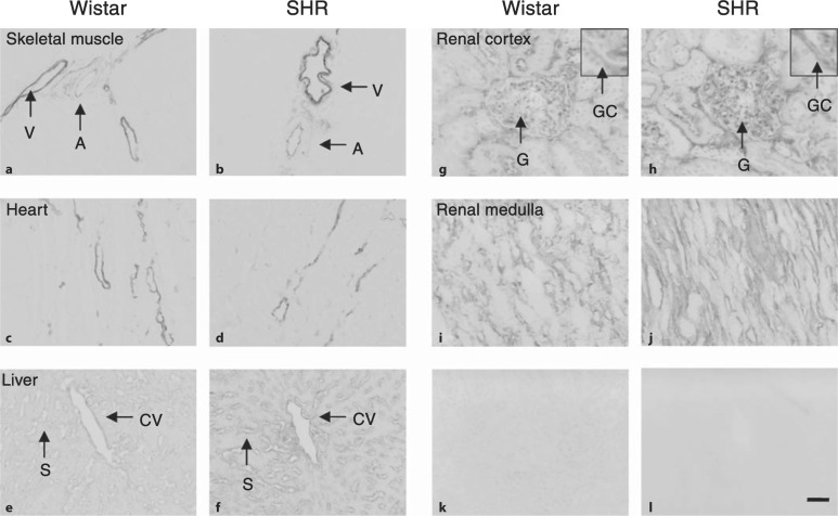
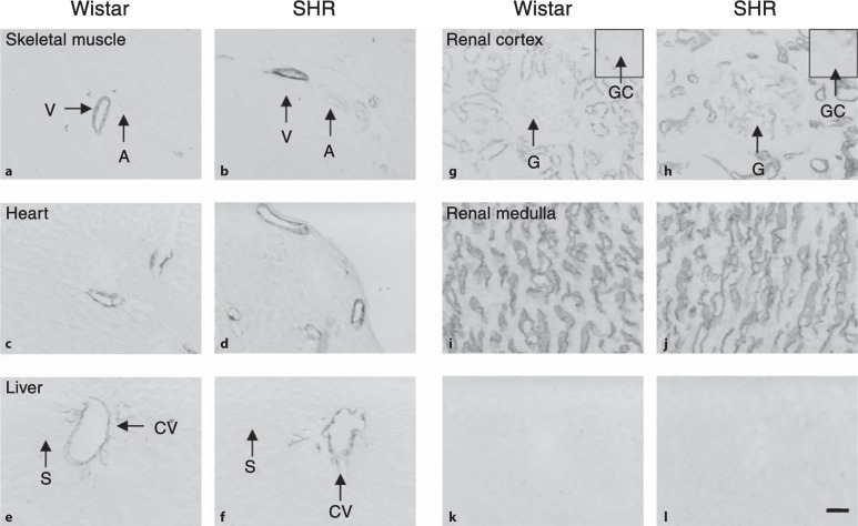
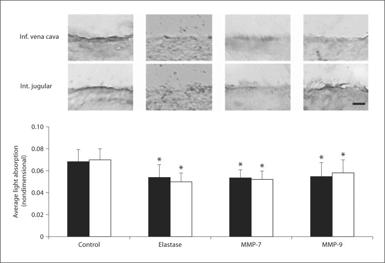
Methods: ICAM-1 was labeled on tissue sections with two antibodies targeting its extracellular and intracellular domains and evaluated by light absorption measurements. The in situ cleavage of ICAM-1 was assessed by treating vessel sections with matrix metalloproteinase (MMP)-7, MMP-9 and elastase.

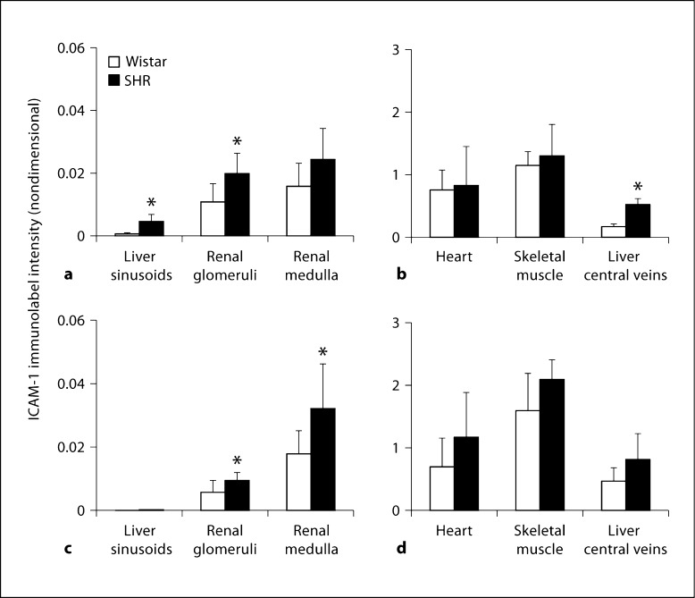
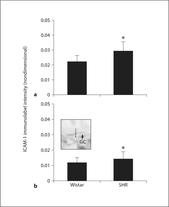
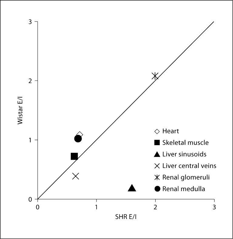
Results: SHRs showed a significant increase in ICAM-1 expression in liver and kidney compared with Wistar rats. The liver and kidney glomeruli exhibit a discrepancy in label density between intra- and extracellular antibodies, which suggests that enzymatic cleavage may be a factor determining ICAM-1 distribution. MMP-7 and MMP-9, which are elevated in SHR plasma, and elastase, which has elevated activity in SHR neutrophils, cleave the extracellular domain of ICAM-1 when applied to the tissue.

Conclusion: ICAM-1 expression in SHRs is upregulated in a tissue-specific manner. Proteolytic cleavage of the extracellular domain of ICAM-1 and accumulation in kidney glomeruli may play a role in the renal involvement of inflammation.


Figures

Fig. 1
Fig. 1
Micrographs of selected tissue cryosections after labeling with an antibody against the extracellular domain of ICAM-1 (1A29). k, l are negative control sections of renal cortex and renal medulla, respectively, after replacement of primary antibody with control mouse IgG. Negative controls of other tissues have similar label intensity. A = Artery; V = vein; CV = liver central vein; S = liver sinusoid; G = renal glomeruli; GC = glomerular capsule. The scale bar represents 50 μm.
Fig. 2
Fig. 2
Micrographs of selected tissue cryosections after labeling with an antibody against the intracellular domain of ICAM-1 (M-19). k, l are negative control sections of renal cortex and renal medulla, respectively, after replacement of primary antibody with control goat IgG. Negative controls of other tissues have similar label intensity. A = Artery; V = vein; CV = liver central vein; S = liver sinusoid; G = renal glomeruli; GC = glomerular capsule. The scale bar represents 50 μm.
Fig. 3
Fig. 3
Quantitative analysis of ICAM-1 label intensity. ICAM-1 label intensity on tissue sections labeled with the extracellular domain antibody 1A29 (a, b) and the intracellular domain antibody M-19 (c, d) is evaluated by light absorption, calculated as described in the Material and Methods section. The number of animals per group was 6 for renal cortex and medulla and 3 elsewhere. ∗ p < 0.05 versus Wistar.
Fig. 4
Fig. 4
Quantitative analysis of ICAM-1 label intensity on renal glomerular capsules (GC). ICAM-1 label intensity was calculated at selected cross-sections along each glomerular capsule (dotted line in inset). a Glomerular capsules labeled with the extracellular domain antibody 1A29. b Glomerular capsules labeled with the intracellular domain antibody M-19. The number of animals per group was 6. ∗ p < 0.05 versus Wistar.
Fig. 5
Fig. 5
Plasma concentration of soluble ICAM-1. ∗ p < 0.05 versus Wistar.
Fig. 6
Fig. 6
Enzymatic cleavage of ICAM-1. Sections of inferior vena cava (▪) and internal jugular vein (□) were incubated with PBS, elastase, MMP-7 and MMP-9. After incubation, the sections were stained with an antibody against the extracellular domain of ICAM-1 (1A29). Top: micrographs of tissue sections; bottom: quantitative analysis of ICAM-1 label intensity. ∗ p < 0.05 versus control. The scale bar represents 10 μm.
Fig. 7
Fig. 7
Elastase activity of blood. a Elastase activity of neutrophils. Insets: fluorescence images of neutrophils after treatment with a fluorogenic elastase substrate. b Elastase activity of plasma. ∗ p < 0.05 versus Wistar; + p < 0.05 versus control. The scale bar represents 5 μm.
Fig. 8
Fig. 8
Cross-correlation of the ratio between average extra- and intra-cellular label density of ICAM-1 (E/I) in different Wistar and SHR tissues. Each point represents the average E/I ratio (the label intensity of 1A29 divided by that of M-19) for one tissue of SHRs and Wistar rats. The line of identity represents equal labeling ratios in the two strains.

Similar articles

Cited by

References

    1. Hilgers KF. Monocytes/macrophages in hypertension. J Hypertens. 2002;20:593–596. - PubMed
    1. Suematsu M, Suzuki H, Delano FA, Schmid-Schönbein GW. The inflammatory aspect of the microcirculation in hypertension: oxidative stress, leukocytes/endothelial interaction, apoptosis. Microcirculation. 2002;9:259–276. - PubMed
    1. Guzik TJ, Hoch NE, Brown KA, McCann LA, Rahman A, Dikalov S, Goronzy J, Weyand C, Harrison DG. Role of the T cell in the genesis of angiotensin II induced hypertension and vascular dysfunction. J Exp Med. 2007;204:2449–2460. - PMC - PubMed
    1. Clozel M, Kuhn H, Hefti F, Baumgartner HR. Endothelial dysfunction and subendothelial monocyte macrophages in hypertension. Effect of angiotensin converting enzyme inhibition. Hypertension. 1991;18:132–141. - PubMed
    1. Rodriguez-Iturbe B, Quiroz Y, Ferrebuz A, Parra G, Vaziri ND. Evolution of renal interstitial inflammation and NF-κB activation in spontaneously hypertensive rats. Am J Nephrol. 2004;24:587–594. - PubMed

Publication types

MeSH terms

Substances