. 2011 May 31;43(5):281-90.
doi: 10.3858/emm.2011.43.5.029.

Introduction of the CIITA gene into tumor cells produces exosomes with enhanced anti-tumor effects

Affiliations

Introduction of the CIITA gene into tumor cells produces exosomes with enhanced anti-tumor effects

Yeong Shin Lee et al. Exp Mol Med. .

Abstract

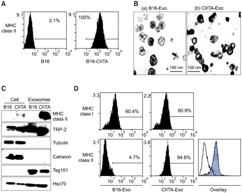
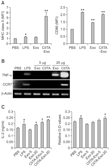
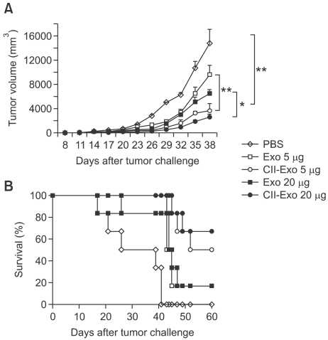
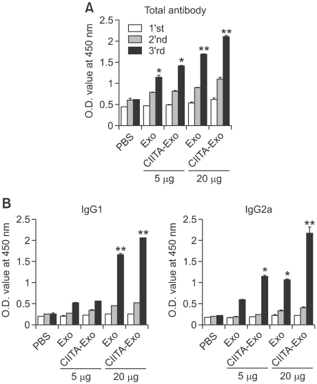
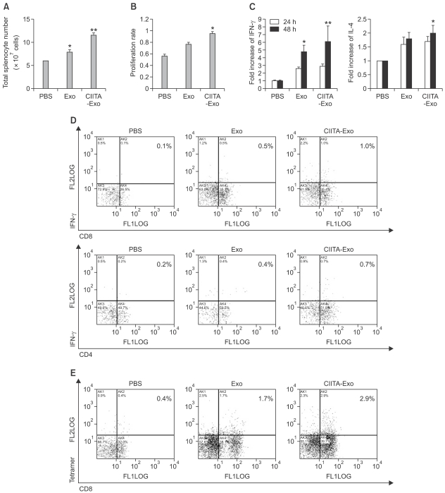
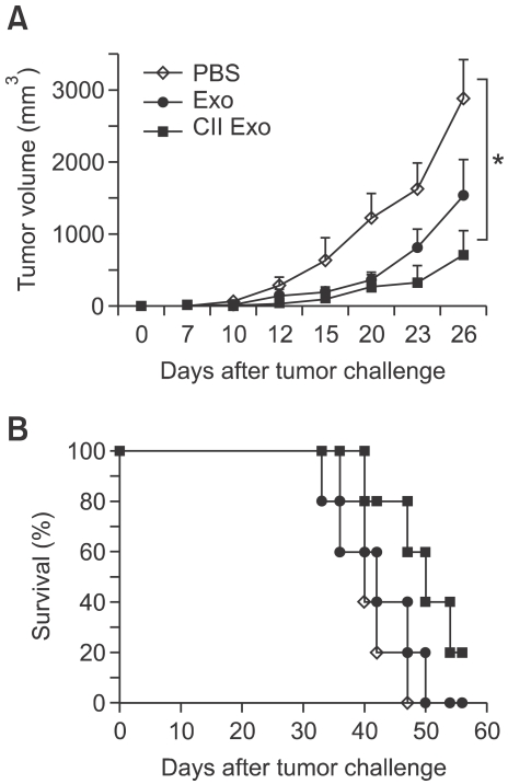
Exosomes are small membrane vesicles secreted from various types of cells. Tumor-derived exosomes contain MHC class I molecules and tumor-specific antigens, receiving attention as a potential cancer vaccine. For induction of efficient anti-tumor immunity, CD4+ helper T cells are required, which recognize appropriate MHC class II-peptide complexes. In this study, we have established an MHC class II molecule-expressing B16F1 murine melanoma cell line (B16F1- CIITA) by transduction of the CIITA (Class II transactivator) gene. Exosomes from B16-CII cells (CIITA- Exo) contained a high amount of MHC class II as well as a tumor antigen TRP2. When loaded on dendritic cells (DCs), CIITA-Exo induced the increased expression of MHC class II molecules and CD86 than the exosomes from the parental cells (Exo). In vitro assays using co-culture of immunized splenocytes and exosome-loaded DCs demonstrated that CIITA-Exo enhanced the splenocyte proliferation and IL-2 secretion. Consistently, compared to B16-Exo, CIITA-Exo induced the increased mRNA levels of inflammatory cytokines such as TNF-α, chemokine receptor CCR7 and the production of Th1-polarizing cytokine IL-12. A tumor preventive model showed that CIITA-Exo significantly inhibited tumor growth in a dose-dependent manner. Ex vivo assays using immunized mice demonstrated that CIITA-Exo induced a higher amount of Th1-polarized immune responses such as Th1-type IgG2a antibodies and IFN-γ cytokine as well as TRP2-specific CD8+ T cells. A tumor therapeutic model delayed effects of tumor growth by CIITA-Exo. These findings indicate that CIITA-Exo are more efficient as compared to parental Exo to induce anti-tumor immune responses, suggesting a potential role of MHC class II-containing tumor exosomes as an efficient cancer vaccine.


Figures

Figure 1
Figure 1
Characterization of exosomes from CIITA-transducted tumor cells. (A). MHC class II expression on CIITA-transfected B16F1 tumor cells. The clone cell were analysed by flow cytometry to evaluation expression of MHC class II molecules. (B) exosomes were observed by electron microscopy (Bar=100 nm) (C) cellular proteins and exosomes from mock (B16) or CIITA transduced (CIITA) cells were analyzed by immunoblot analysis with each indicated antibodies against MHC class II, TRP-2, Tsg101, Hsc70, tubulin, and Calnexin. (D) Bead-exosomes complex were incubated with PE-conjugated anti-MHC class I and II antibodies, and analyzed by flow cytometry.
Figure 2
Figure 2
Immune stimulating effects of CIITA-Exo on dendtitic cell line. (A) The surface expression of MHC class II and CD86 on the pulsed DCs was detected by flow cytometry. The number above the bars indicates the fold increase of mean fluorescence intensity (MFI) (B) The mRNA level of TNF-a, CCR-7 and β-actin in the pulsed DCs was analyzed by RT-PCR. (C) The culture supernatants were collected to determine IL-2 release by ELISA and CTLL-2D proliferation assay (alarmar blue). Statistical significance, *: P <0.05, ** : P <0.005.
Figure 3
Figure 3
More potent protective antitumor immunity induced by immunization with CIITA-Exo in preventive tumor models. C57BL/6 mice (n=7 per group) were intradermally immunized with PBS (◊), Exo 5 µg (□), Exo 20 µg (■), CIITA-Exo 5 µg (○) or CIITA-Exo 20 µg (●). One week after the last immunization, the immunized mice were challenged with B16F1 tumor cells. (A) Tumor volume was measured every three days from one week after tumor challenge, and was calculated using the formula longer length × shorter length2/2. (B) Survival of each group of mice was monitored until two months after tumor challenge. Statistical significance, *: P <0.05, ** : P <0.005.
Figure 4
Figure 4
Humoral immune responses by exosomes were analyzed. Mice were immunized three times with either PBS, Exo 5 µg or Exo 20 µg or CIITA-Exo 5 µg or CIITA-Exo 20 µg. The experiments were repeated twice and displayed similar results. Sera from the immunized mice were obtained and were pooled one week after each immunization (1st, 2nd and 3rd week). (A) Anti-mouse IgG and IgM antibody was used to detect whole antibodies. (B) Anti-IgG1- and anti-IgG2a-speicific antibodies were used to detect antibody isotypes. Statistical significance; ns: not significant, *: P <0.05, **: P <0.01.
Figure 5
Figure 5
Cellular immune responses by exosomes were analyzed. Mice were immunized three times with either PBS, Exo 5 µg or Exo 20 µg or CIITA-Exo 5 µg or CIITA-Exo 20 µg. Spleens from the respective groups were extracted 10 days after the last immunization. (A) The absolute cell numbers of splenocytes were counted by trypan blue exclusion assay. (B) One week after the last immunization, splenocytes from the respective groups were harvested and pooled together. Equal numbers of splenocytes from respective immunized mouse groups were stimulated with mitomycin C-treated B16 cells. After 72 hr stimulation, the stimulated splenocytes were assayed by alarma blue assay for cell proliferation. The experiments were performed twice and demonstrated similar result. Statical significance; *P <0.05. (C) The co-culture supernatnats were collected after 24 or 48 h incubation and utilized for ELISA to measure the quantity of IFN-γ or IL-4 secreted from the stimulated splenocytes. *P <0.05. (D) Intrcellular cytocine assay. The stimulated splenocytes were surface-stained with FITC anti CD-4 or anti CD-8 antibodies and intracelluarly labeled with PE anti-IFN-γ or anti-IL-4 antibodies. (E) Tetramer assay. The stimulated splenocytes were stained with FITC-tagged anti-CD8 antibodies and were reacted with PE-conjugated H-2b/TRP2 tetramer. The percentage of the double-positive cell population is displayed on each panel.
Figure 6
Figure 6
In vivo anti-tumor effects of Exo in tumor-bearing mouse were evaluated. C57BL/6 mice (n = 7 per group) were injected with B16F1 tumor cells. When the tumor size reached 1 cm2, either PBS (◊), Exo 20 µg (●) or CIITA-Exo 20 µg (■) was intradermally injected three times with a two-week interval in therapeutic tumor models. (A) Tumor size was measured one or two times a week. (B) The survival rate of the mice was determined until 60 days after the last injection. Representative data are shown from two repeated independent experiments. Statistical significance; *: P <0.05.

Similar articles

Cited by

References

    1. André F, Chaput N, Schartz NE, Flament C, Aubert N, Bernard J, et al. Exosomes as potent cell-free peptide-based vaccine. I. Dendritic cell-derived exosomes transfer functional MHC class I/peptide complexes to dendritic cells. J Immunol. 2004;172:2126–2136. - PubMed
    1. André F, Schartz NE, Chaput N, Flament C, Raposo G, Amigorena S, Angevin E, Zitvogel L. Tumor-derived exosomes: a new source of tumor rejection antigens. Vaccine. 2002;(Suppl 4):A28–A31. - PubMed
    1. Andre F, Schartz NE, Movassagh M, Flament C, Pautier P, Morice P, et al. Malignant effusions and immunogenic tumour-derived exosomes. Lancet. 2002;360:295–305. - PubMed
    1. Armstrong TD, Clements VK, Martin BK, Ting JP, Ostrand-Rosenberg S. Major histocompatibility complex class II-transfected tumor cells present endogenous antigen and are potent inducers of tumor-specific immunity. Proc Natl Acad Sci USA. 1997;94:6886–6891. - PMC - PubMed
    1. Baxevanis CN, Voutsas IF, Tsitsilonis OE, Gritzapis AD, Sotiriadou R, Papamichail M. Tumor-specific CD4+ T lymphocytes from cancer patients are required for optimal induction of cytotoxic T cells against the autologous tumor. J Immunol. 2000;164:3902–3912. - PubMed

Publication types

MeSH terms

LinkOut - more resources