. 2011 Mar 2;6(1):1-9.
doi: 10.6026/97320630006001.

Structural inferences for Cholera toxin mutations in Vibrio cholerae

Structural inferences for Cholera toxin mutations in Vibrio cholerae

Gunasagaran Shamini et al. Bioinformation. .

Abstract

Cholera is a global disease that has persisted for millennia. The cholera toxin (CT) from Vibrio cholerae is responsible for the clinical symptoms of cholera. This toxin is a hetero-hexamer (AB(5)) complex consisting of a subunit A (CTA) with a pentamer (B(5)) of subunit B (CTB). The importance of the AB(5) complex for pathogenesis is established for the wild type O1 serogroup using known structural and functional data. However, its role is not yet documented in other known serogroups harboring sequence level residue mutations. The sequences for the toxin from different serogroups are available in GenBank (release 177). Sequence analysis reveals mutations at several sequence positions in the toxin across serogroups. Therefore, it is of interest to locate the position of these mutations in the AB(5) structure to infer complex assembly for its functional role in different serogroups. We show that mutations in the CTA are at the solvent exposed regions of the AB(5) complex, whereas those in the CTB are at the CTB/CTB interface of the homo-pentamer complex. Thus, the role of mutations at the CTB/CTB interface for B(5) complex assembly is implied. It is observed that these mutations are often non-synonymous (e.g. polar to non-polar or vice versa). The formation of the AB(5) complex involves inter-subunit residue-residue interactions at the protein-protein interfaces. Hence, these mutations, at the structurally relevant positions, are of importance for the understanding of pathogenesis by several serogroups. This is also of significance in the improvement of recombinant CT protein complex analogs for vaccine design and their use against multiple serogroups.

Keywords: Cholera toxin (CT); O1/O139; Vibrio cholerae; mutation; non O1/O139; protein-protein interfaces.


Figures

Figure 1
Figure 1
Structural model of a cholera toxin (CT). CT is a hetero-hexameric complex (AB5) consisting of CTA (194 residues A1 and 46 residues A2) and CTB (103 residues) pentamer with D, E, F, G and H chains.
Figure 2
Figure 2
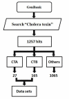
Creation of sequence dataset for CTA and CTB. A sequence dataset of CTA and CTB was derived from GenBank (release 177) using KEYWORD search as illustrated in the flowchart. The KEYWORD search “cholera toxin” resulted in 1257 hits. This set consists of 27 CTA sequences, 165 CTB sequences according to GenBank description and available annotations. The remaining 1065 sequences with descriptions such as secretion protein, cholera toxin transcriptional activator, ADP-ribosylation factor, GNAS complex, dopamine receptor, Pertusis toxin, Shiga-like toxin and the like are eliminated from the dataset. Thus, a CT sequence dataset of 192 sequences (Table 1 in supplementary material) consisting of 27 CTA and 165 CTB was created. The CTA and CTB sequences are included in the dataset as available in the GenBank. The biased availability on the amount of CTA and CTB sequences in GenBank is attributed to the likely observation of frequent mutations in CTB.
Figure 3
Figure 3
MSA for the CTA subunit of different serogroups. The MSA was performed using the wild type O1 classical strain sequence with known structure (PDB ID: 1XTC) as reference. The position specific mutations among the available CTA sequences (27) with reference to the classical sequence are indicated using dark shades. CT is an AB5 hetero-hexamer and hence, the CTA/CTB interface residues in CTA are indicated using light shades.
Figure 4
Figure 4
MSA for the CTB subunit of different serogroups. The MSA was performed using the wild type O1 Classical strain with known structure (PDB ID:1XTC) as reference. The position specific mutations among the available CTB sequences (165) with reference to the Classical sequence are indicated using dark shades. B5 is a homo-pentamer and hence, the CTB/CTB interface residues in B5 are indicated using light shades. It should be noted that the mutated residues at the CTB/CTB interfaces in B5 are highlighted using both dark and light shades at their corresponding position specific residues.
Figure 5
Figure 5
Structural model of CTB/CTB interfaces in B5. B5 is a homopentamer and each CTB subunit (D) is juxtaposed by two other CTB units on either side (E and H). Thus, the D subunit creates two different types of interfaces (D-E and D-H) on either side. This subsequently results in two different “position specific interacting” patterns in sequence for subunit D.
Figure 6
Figure 6
Representation of mutated residue positions in serogroups to interface residues in CT complex as a function of their residue position identified using ΔASA measure.
  1. Mapping of CTA mutations to CTA/CTB interface residues in CTA (Please refer to Figure 1 for the visual illustration of CTA/CTB interface).

  2. Mapping of CTB mutations to CTB (D subunit)/CTB (E subunit) interface residues (Please refer to Figure 5 for the visual illustration of D-E interface).

  3. Mapping of CTB mutations to CTB (D subunit)/CTB (H subunit) interface residues (Please refer to Figure 5 for the visual illustration of D-H interface).

It should be noted that mutated residue positions are mapped on to corresponding interface residue positions in all the three cases (a), (b) and (c).
Figure 7
Figure 7
Structural models of CTA (a) and CTB (b) subunits with known mutations among archived serogroups. We used the structure with PDB entry (1XTC) for generating this visual using the freeware Discovery studio from Accelrys Inc.
  1. A total of 6 unique mutations thus observed among the known CTA sequences (Table 2 in supplementary material) from several serogroups are shown at their corresponding 6 residue positions using the Corey-Pauling-Kultun (CPK) residue model representation.

  2. Fourteen unique mutations thus observed among the known CTB sequences (Table 3 in supplementary material) from several serogroups are shown at their corresponding 13 residue positions using the CPK residue model representation.

Figure 8
Figure 8
Structural models of CTA (a) and CTB (b) subunits with known mutations at the respective structural interfaces or solvent accessible regions in the complex among archived serogroups. We used the structure with PDB entry (1XTC) for generating this visual using the freeware Discovery studio from Accelrys Inc.
  1. A total of 6 unique mutations thus observed among the known CTA sequences (Table 4 in supplementary material) from several serogroups are shown at their corresponding 6 residue positions using the CPK residue model representation. All of these 6 mutated positions are present at the solvent exposed regions of CTA in both monomer and CTA/CTB complex state.

  2. A total of 7 out of 14 unique mutations thus observed among the known CTB sequences (Table 4 in supplementary material) from several serogroups are shown at their corresponding 7 (3, 15, 25, 34, 47,52 and 60) out of the 13 residue positions using the CPK residue model representation are at the CTB/CTB interfaces in the B5 complex.

References

    1. WHO Cholera 2009. Wkly Epidemiol Rec. 2010;85:293.
    1. Centers for Disease Control and Prevention (CDC) Update: Cholera Outbreak - Haiti, 2010. MMWR Morb Mortal Wkly Rep. 2010;59:1473. - PubMed
    1. T Shimada, et al. Curr Microbiol. 1994;28:175.
    1. SK Bhattarcharya, et al. J Infect. 1993;27:11. - PubMed
    1. JB Kaper, et al. Clin Microbiol Rev. 1995;8:48. - PMC - PubMed

LinkOut - more resources