. 2011 Mar 25;6(3):e18065.
doi: 10.1371/journal.pone.0018065.

Mitotic defects lead to pervasive aneuploidy and accompany loss of RB1 activity in mouse LmnaDhe dermal fibroblasts

Affiliations

Mitotic defects lead to pervasive aneuploidy and accompany loss of RB1 activity in mouse LmnaDhe dermal fibroblasts

C Herbert Pratt et al. PLoS One. .

Abstract

Background: Lamin A (LMNA) is a component of the nuclear lamina and is mutated in several human diseases, including Emery-Dreifuss muscular dystrophy (EDMD; OMIM ID# 181350) and the premature aging syndrome Hutchinson-Gilford progeria syndrome (HGPS; OMIM ID# 176670). Cells from progeria patients exhibit cell cycle defects in both interphase and mitosis. Mouse models with loss of LMNA function have reduced Retinoblastoma protein (RB1) activity, leading to aberrant cell cycle control in interphase, but how mitosis is affected by LMNA is not well understood.

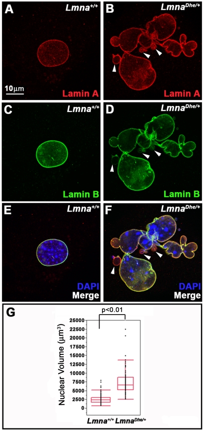
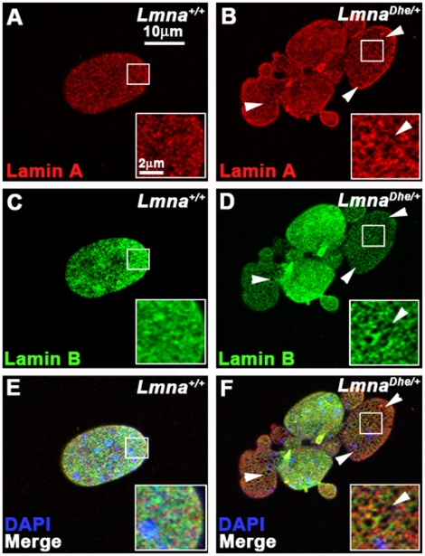
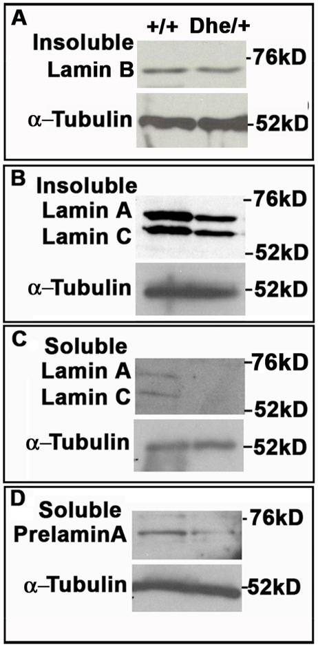
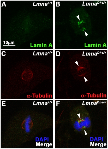
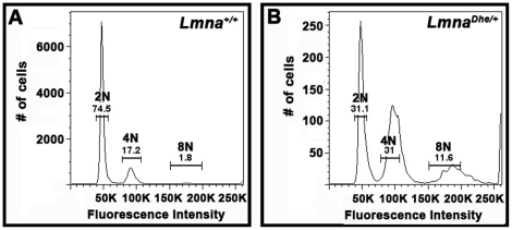
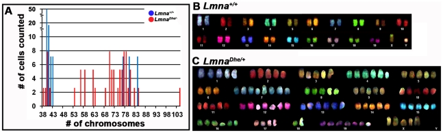
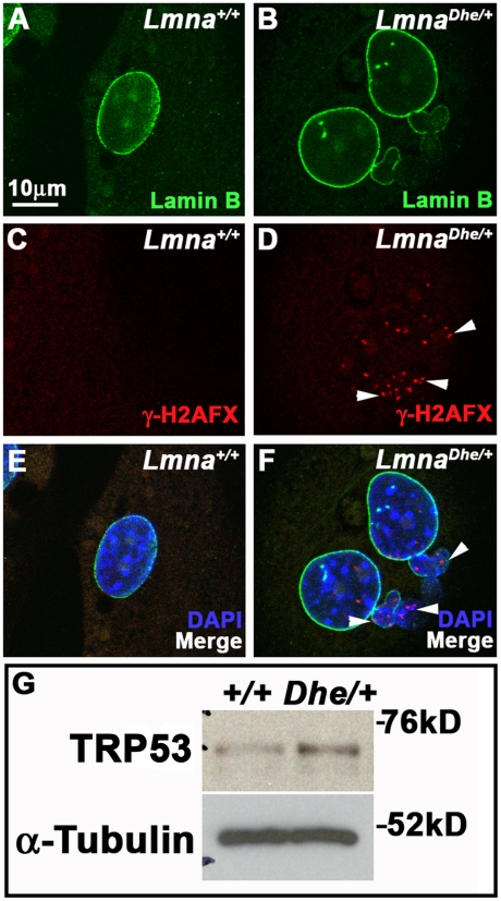
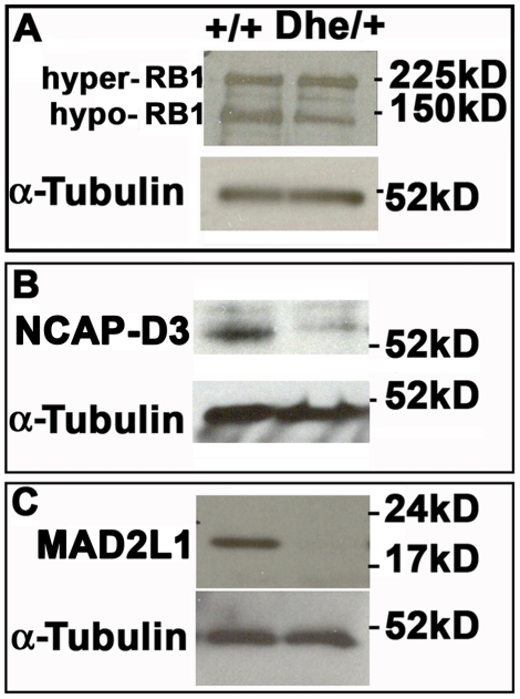
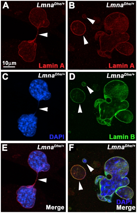
Results: We examined the cell cycle and structural phenotypes of cells from mice with the Lmna allele, Disheveled hair and ears (Lmna(Dhe)). We found that dermal fibroblasts from heterozygous Lmna(Dhe) (Lmna(Dhe/+)) mice exhibit many phenotypes of human laminopathy cells. These include severe perturbations to the nuclear shape and lamina, increased DNA damage, and slow growth rates due to mitotic delay. Interestingly, Lmna(Dhe/+) fibroblasts also had reduced levels of hypophosphorylated RB1 and the non-SMC condensin II-subunit D3 (NCAP-D3), a mitosis specific centromere condensin subunit that depends on RB1 activity. Mitotic check point control by mitotic arrest deficient-like 1 (MAD2L1) also was perturbed in Lmna(Dhe/+) cells. Lmna(Dhe/+) fibroblasts were consistently aneuploid and had higher levels of micronuclei and anaphase bridges than normal fibroblasts, consistent with chromosome segregation defects.

Conclusions: These data indicate that RB1 may be a key regulator of cellular phenotype in laminopathy-related cells, and suggest that the effects of LMNA on RB1 include both interphase and mitotic cell cycle control.


Conflict of interest statement

Competing Interests: The authors have declared that no competing interests exist.

Figures

Figure 1
Figure 1. LmnaDhe/+ cells exhibit blebbed nuclear membranes and large nuclear volumes.
(A–F) Immunodetection of Lamin A (red; A,B, E,F) and Lamin B (green; C–F) in Lmna+/+(A,C,E) and LmnaDhe/+ (B,D,F) fibroblasts reveals extensive blebbing of the nuclear envelope and exclusion of Lamin B from these blebs in mutant cells (white arrowheads in B,D,F). Single confocal optical sections through the middle of the nucleus, counterstained with DAPI (blue), are shown. (G) Box-and-whisker plot of nuclear volumes measured from 3D confocal image stacks indicates a significantly greater average volume and greater range in nuclear sizes in LmnaDhe/+ versus Lmna+/+ fibroblasts (p<0.01; Student's t-test).
Figure 2
Figure 2. LMNADhe affects both the Lamin A and Lamin B meshwork.
(A–F) Immunodetection of Lamin A (red; A,B,E,F) and Lamin B (green;C–F) in Lmna+/+(A,C,E) and LmnaDhe/+ (B,D,F) fibroblasts reveals large, irregular holes in the Lamin A (white arrowheads in B) and Lamin B meshwork (white arrows in D) in mutant cells. Insets represent magnified portions of an area of the nucleus indicted by the white boxes. Single confocal optical sections through the top of the nucleus are shown. (E, F) Merged images with nuclei counterstained with DAPI (blue).
Figure 3
Figure 3. LmnaDhe/+ fibroblasts exhibit lower Lamin A levels, but no change in Lamin B expression.
(A–D) Western blot analysis of insoluble Lamin B (A), insoluble Lamin A (B), soluble Lamin A (C) and soluble Prelamin A (D). These data indicate no difference in Lamin B protein levels between Lmna+/+and mutant cells, but less soluble Lamin A/C, insoluble Lamin A/C and Prelamin A protein in mutant cells. α-Tubulin was used as a loading control for all western blots.
Figure 4
Figure 4. LMNA persists and colocalizes with the spindle apparatus during metaphase in LmnaDhe/+ cells.
(A–F) Immunodetection of Lamin A (green;A,B,E,F) and α-Tubulin (red; C–F) in Lmna+/+(A,C,E) and LmnaDhe/+ (B,D,F) fibroblasts. Labeling indicates persistence of Lamin A in mitotic cells and colocalization with α-Tubulin during metaphase of mutant cells (white arrowheads), as opposed to Lmna+/+cells. Single confocal optical sections through the middle of the cell are shown. The metaphase chromosomes are counterstained with DAPI (blue: E, F).
Figure 5
Figure 5. LmnaDhe/+ fibroblasts have increased DNA content subsequent to pervasive aneuploidy.
(A and B) Flow cytometry analysis of propidium iodide labeling of Lmna+/+(A) and LmnaDhe/+ (B) dermal fibroblasts indicates that mutant cells have a significantly higher proportion of cells with 4C and 8C and lower fraction of 2C cells, as compared to Lmna+/+cell populations (p<0.01, Student's t-test). DNA content peaks for LmnaDhe/+ were also broader than Lmna+/+peaks, indicating a population of cells with a large range of DNA content.
Figure 6
Figure 6. Spectral karyotyping reveals pervasive aneuploidy in LmnaDhe/+ fibroblasts.
(A) Histogram of chromosome numbers in LmnaDhe/+ (red bars) and Lmna+/+ (blue bars) dermal fibroblasts, as determined by metaphase spreading and SKY. Over 90% of mutant cells possessed an abnormal number of chromosomes ranging from 38 to 104. (B and C) Representative spectral karyotypes of Lmna+/+ (B) and LmnaDhe/+ (C) cells. The wild-type cell exhibited a normal karyotype of 40XY (B), while the mutant cell was aneuploid with a 104XXXXX karyotype (C).
Figure 7
Figure 7. LmnaDhe/+ fibroblasts have foci of γH2AX and increased levels of TRP53 indicating DNA damage.
(A–F) Immunodetection of Lamin B (green; A,B, E,F) and γH2AX (red; C–F) in Lmna+/+(A,C,E) and LmnaDhe/+ (B,D,F) fibroblasts. Mutant fibroblast nuclei have several foci of γH2AX throughout both the blebbed and non-blebbed areas of the nucleus (white arrowheads in D and F), in contrast to Lmna+/+nuclei. Single confocal optical sections though the middle of the nucleus are shown. Nuclei are counterstained with DAPI. (G) Western blots of total TRP53 in mutant and Lmna+/+fibroblasts show higher levels of TRP53 in mutant cells than in Lmna+/+ cells. α-Tubulin was used as loading control.
Figure 8
Figure 8. Decreased levels of hypophosphorylated RB1, NCAP-D3 and MAD2L1 in LmnaDhe/+ fibroblasts.
(A) Western blotting indicated a lower level of activated, hypo-phosphorylated RB1 (150 kD band) in mutant cells, while hyper-phosphorylated RB1 levels were slightly elevated (225 kD band). (B) LmnaDhe/+ fibroblasts express lower levels of the centromere-specific cohesin subunit, NCAP-D3, as well. (C) LmnaDhe/+ cells express little to no detectable MAD2L1, a spindle checkpoint protein, while Lmna+/+cells robustly expressed this protein. α-Tubulin was used as a loading control for all western blots.
Figure 9
Figure 9. Increased occurrence of micronuclei and anaphase bridges in LmnaDhe/+ fibroblasts indicate segregation defects during mitosis.
(A–F) Immunodetection of Lamin A (red; A,B,E,F) and Lamin B (green; C–F) in LmnaDhe/+ fibroblasts. Mutant fibroblast cultures have an increased proportion of cells with anaphase bridges (A,C,E; white arrowheads) (Table 3; p<0.001; t-test) and micronuclei (B,D,F; white arrowheads) (Table 3; p<0.001; t-test), as compared to Lmna+/+ cells, consistent with chromosome segregation defects during mitosis. Single confocal optical sections though the middle of the nucleus are shown. Nuclei are counterstained with DAPI.

References

    1. Worman HJ, Fong LG, Muchir A, Young SG. Laminopathies and the long strange trip from basic cell biology to therapy. J Clin Invest. 2009;119:1825–1836. - PMC - PubMed
    1. Worman HJ, Bonne G. “Laminopathies”: a wide spectrum of human diseases. Exp Cell Res. 2007;313:2121–2133. - PMC - PubMed
    1. Stewart CL, Kozlov S, Fong LG, Young SG. Mouse models of the laminopathies. Exp Cell Res. 2007;313:2144–2156. - PMC - PubMed
    1. Shimi T, Pfleghaar K, Kojima S, Pack CG, Solovei I, et al. The A- and B-type nuclear lamin networks: microdomains involved in chromatin organization and transcription. Genes Dev. 2008;22:3409–3421. - PMC - PubMed
    1. Dechat T, Pfleghaar K, Sengupta K, Shimi T, Shumaker DK, et al. Nuclear lamins: major factors in the structural organization and function of the nucleus and chromatin. Genes Dev. 2008;22:832–853. - PMC - PubMed

Publication types

MeSH terms

Associated data