Review
. 2011 Jul;20 Suppl 2(Suppl 2):S330-5.
doi: 10.1007/s00586-011-1783-9. Epub 2011 Apr 6.

Intradural extramedullary spinal inflammatory myofibroblastic tumor: case report and literature review

Affiliations
Review

Intradural extramedullary spinal inflammatory myofibroblastic tumor: case report and literature review

Ilyess Zemmoura et al. Eur Spine J. 2011 Jul.

Abstract

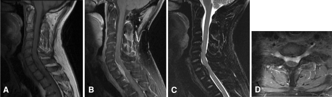
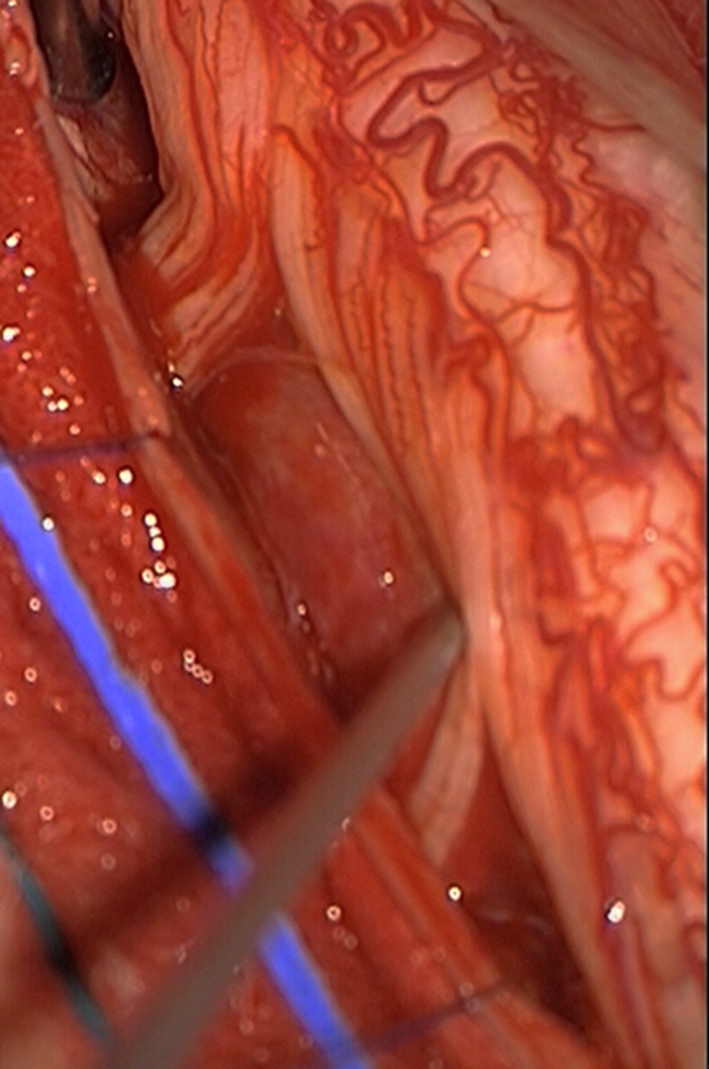
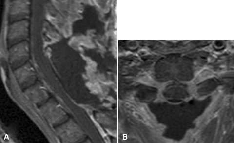
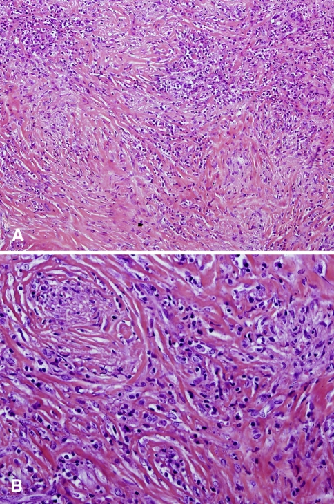
The authors present the case of an inflammatory myofibroblastic tumor that involves the cervical spinal cord meninges, presenting in a manner mimicking en plaque meningioma, which has never been previously reported. During the first surgical procedure, which did not involve exploration of the intradural space, inflammatory epidural tissue was found. We performed a second operation to remove the tumor that was finally intradural, dural-based and very tough. Imaging studies, surgical findings, and histopathological examinations were used to support the diagnosis. Intradural extramedullary inflammatory myofibroblastic tumor is a rare entity that has only been described nine times in the literature. Surgery remains the treatment of choice. Although histologically benign, spinal inflammatory myofibroblastic tumor can be aggressive and requires a large resection and long-term follow-up of the entire central nervous system with magnetic resonance imaging.


Figures

Fig. 1
Fig. 1
Preoperative magnetic resonance imaging (MRI). a Sagittal T1-weighted image showing a tumor isointense to the spinal cord, at C6 to T1. b Homogeneous enhancement of the tumor after injection of gadolinium. c Sagittal T2-weighted image: the tumor appears as very low signal intensity. d Axial post-contrast T1-weighted image: bilateral foraminal contrast enhancement interpreted as extension of the tumor to the C7 neural foramina
Fig. 2
Fig. 2
Intraoperative photograph. The tumor is located anterior to the spinal cord and nerve roots
Fig. 3
Fig. 3
Postoperative MRI on post-contrast T1-weighted sequences showing complete removal of the tumor. a sagittal image. b axial image
Fig. 4
Fig. 4
Pathology specimen, hematoxylin and eosin. a (original magnification ×100). There are compact fascicular and myxoid areas. b (original magnification ×200). Spindle cells are mixed with numerous lymphocytes and plasma cells

References

    1. Bahadori M, Liebow AA. Plasma cell granulomas of the lung. Cancer. 1973;31:191–208. doi: 10.1002/1097-0142(197301)31:1<191::AID-CNCR2820310127>3.0.CO;2-D. - DOI - PubMed
    1. Fletcher C, Unni KK, Mertens F (2002) Pathology and genetics of tumors of soft tissue and bone. World Health Organization classification of tumors. IARC press, Lyon, France
    1. Boutarbouch M, Arkha Y, Rifi L, Derraz S, El Ouahabi A, El Khamlichi A. Intradural cervical inflammatory pseudotumor mimicking epidural hematoma in a pregnant woman: case report and review of the literature. Surg Neurol. 2008;69:302–305. doi: 10.1016/j.surneu.2006.12.056. - DOI - PubMed
    1. Brandsma D, Jansen GH, Spliet W, Nielen K, Taphoorn MJ. The diagnostic difficulties of meningeal and intracerebral plasma cell granulomas–presentation of three cases. J Neurol. 2003;250:1302–1306. doi: 10.1007/s00415-003-0200-7. - DOI - PubMed
    1. Eimoto T, Yanaka M, Kurosawa M, Ikeya F. Plasma cell granuloma (inflammatory pseudotumor) of the spinal cord meninges: report of a case. Cancer. 1978;41:1929–1936. doi: 10.1002/1097-0142(197805)41:5<1929::AID-CNCR2820410537>3.0.CO;2-S. - DOI - PubMed

MeSH terms