. 2011 Jun;240(6):1402-11.
doi: 10.1002/dvdy.22630. Epub 2011 Apr 4.

Gdnf is mitogenic, neurotrophic, and chemoattractive to enteric neural crest cells in the embryonic colon

Affiliations

Gdnf is mitogenic, neurotrophic, and chemoattractive to enteric neural crest cells in the embryonic colon

Olive Mwizerwa et al. Dev Dyn. 2011 Jun.

Abstract

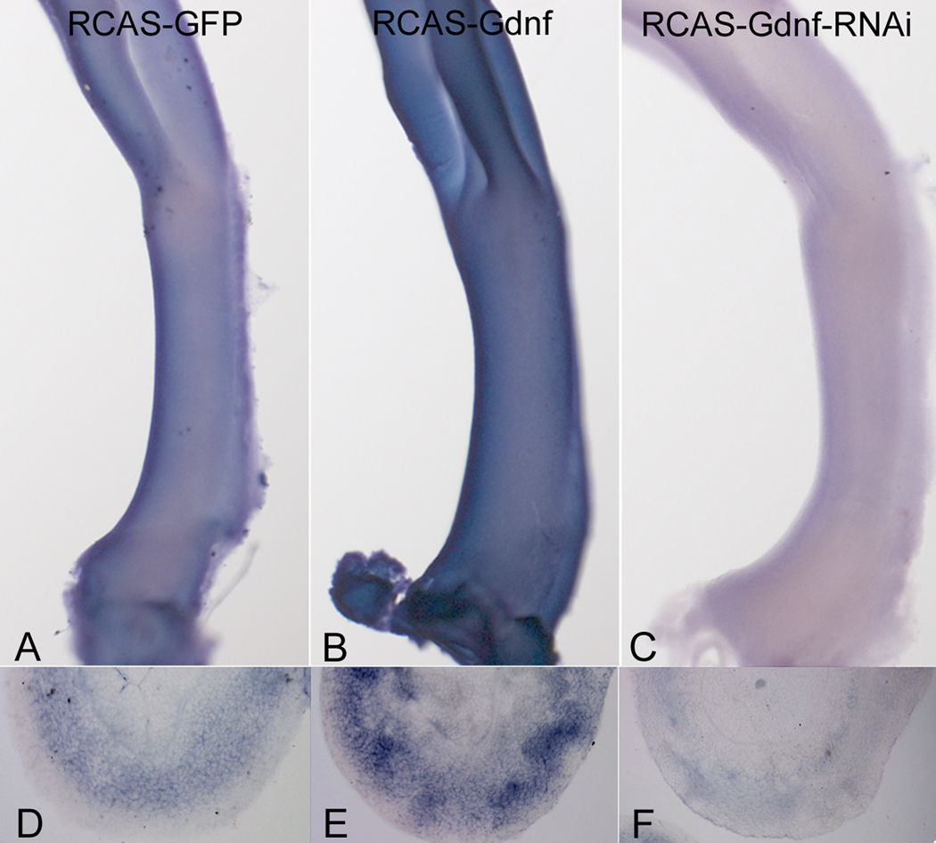
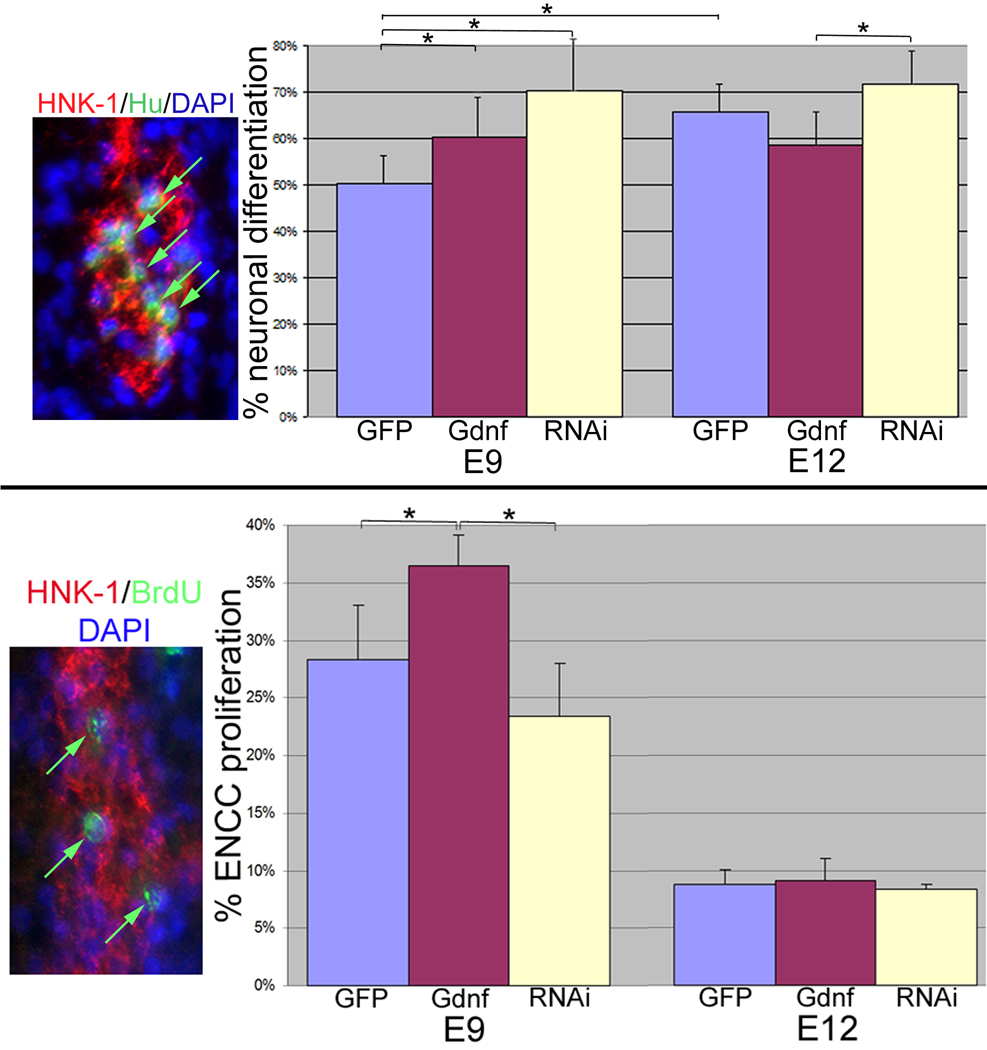
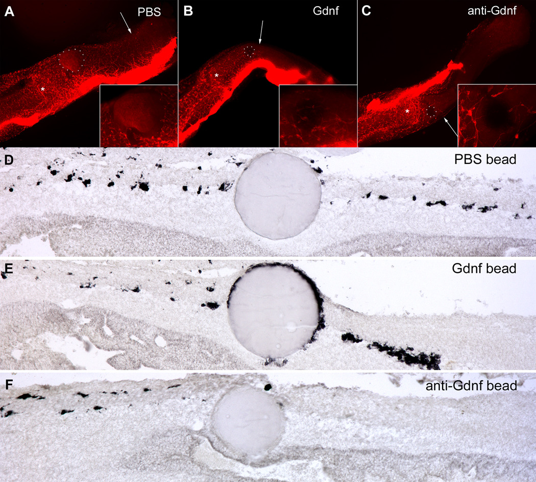
Glial-derived neurotrophic factor (Gdnf) is required for morphogenesis of the enteric nervous system (ENS) and it has been shown to regulate proliferation, differentiation, and survival of cultured enteric neural crest-derived cells (ENCCs). The goal of this study was to investigate its in vivo role in the colon, the site most commonly affected by intestinal neuropathies such as Hirschsprung's disease. Gdnf activity was modulated in ovo in the distal gut of avian embryos using targeted retrovirus-mediated gene overexpression and retroviral vector-based gene silencing. We find that Gdnf has a pleiotropic effect on colonic ENCCs, promoting proliferation, inducing neuronal differentiation, and acting as a chemoattractant. Down-regulating Gdnf similarly induces premature neuronal differentiation, but also inhibits ENCC proliferation, leading to distal colorectal aganglionosis with severe proximal hypoganglionosis. These results indicate an important role for Gdnf signaling in colonic ENS formation and emphasize the critical balance between proliferation and differentiation in the developing ENS.


Figures

Figure 1
Figure 1. Gdnf is required for colonization of the colorectal ENS
E5.5 intestine was cultured in serum-free collagen matrix in the presence (D–F) or absence (A–C) of anti-Gdnf antibody. After 3 days in culture, wholemount immunohistochemistry was performed with Tuj1 (A,B,D,E). The proximal end of the colon is marked with an asterisk and the migratory wavefront with an arrow (A,D). The wavefront region is magnified in B,E. Severe hypoganglionosis is seen throughout the treated colon (D,E), with very sparse and isolated Hu-immunoreactive ganglion cells in cross sections near the wavefront (F). The nerve of Remak (NoR) is smaller in the absence of Gdnf expression (F).
Figure 2
Figure 2. Gdnf expression can be modulated in ovo with RCAS
RCAS virus encoding GFP, Gdnf, or Gdnf-RNAi were injected into the presumptive distal intestinal mesoderm of E2 embryos, and the intestines harvested at E9. Gdnf expression was detected by in situ hybridization on wholemount colorectum (A–C) and on sections through the mid-colon (D–F).
Figure 3
Figure 3. Inhibition of Gdnf produces distal colorectal aganglionosis
Intestines infected with RCAS-GFP (A,C,E,), RCAS-Gdnf-RNAi (B,D,F), or RCAS-Gdnf (G) were collected at E7 (A,B), E8 (C,D), and E9 (E–G). ENS was visualized by wholemount Tuj1 (A–D) or p75NTR (E–G) immunohistochemistry. Distal is to the right in all panels. The proximal end of the colon is marked with an asterisk and the migratory wavefront with an arrow. Insets in A,B are magnified views of the wavefront. Insets in C,D correspond to the proximal colon (1) and the wavefront (2). Magnified views in E-F are from the mid-colon. ENCC migration is markedly delayed in RNAi-infected guts at all stages examined, with the wavefront only reaching the mid-colorectum by E9 (F), when migration is nearly complete in control (E) and RCAS-Gdnf (G) guts. The percent colonization of the colorectum at E8 and E9 is summarized in H, showing a significant delay in ENCC migration in RNAi-infected intestine (*p<0.05 compared to RCAS-GFP and to RCAS-Gdnf).
Figure 4
Figure 4. Gdnf induces premature neuronal differentiation and promotes ENCC proliferation
(Top panel): The percentage of neuronal differentiation was calculated in E9- and E12-injected embryos. Intestine was harvested at those stages and immunohistochemistry performed on cross-sections through the mid-colorectum using antibodies to ENCCs (p75NTR or HNK-1) and neurons (Hu). Panel on left shows a myenteric ganglion from an E12 RCAS-GFP hindgut. The number of neurons and ENCCs was counted to determine the percentage of Hu-immunoreactive ENCCs (arrows). (Bottom panel): ENCC proliferation was determined using BrdU incorporation into HNK-1+ ENCCs. Panel on left shows a myenteric ganglion from an RCAS-GFP infected hindgut with arrows marking BrdU+ cells. The percentage of proliferating ENCCs was calculated by dividing the total number of HNK-1+ ENCCs by the number of BrdU+/HNK-1+ double-immunoreactive cells. The graphs show the average and standard deviation for each group. Lines with asterisk denote statistical significance (p<0.02).
Figure 5
Figure 5. Gdnf is chemoattractive to migrating ENCCs in the colorectum
E5.5 intestine was isolated and the colon implanted with a PBS bead (A,D), Gdnf bead (B,E), or anti-Gdnf bead (C,F), then cultured in collagen gel for 3 days. Wholemount Tuj1 staining shows ENCC migration continuing past the control bead (A), while Gdnf- and anti-Gdnf-coated beads arrested migration (B,C). The proximal end of the colon is marked with an asterisk and the migratory wavefront with an arrow in A–C. Insets show magnified views of the area near the bead. Longitudinal sections (proximal end to the left) were labeled with Hu antibody. ENCCs migrate around the PBS bead (D). In contrast, the Gdnf-coated bead attracts enteric neurons, causing them to cluster around the bead (E). Beads coated with anti-Gdnf antibody stop migration, but do not lead to neuronal clustering (F).

Similar articles

Cited by

References

    1. Amiel J, Lyonnet S. Hirschsprung disease, associated syndromes, and genetics: a review. J Med Genet. 2001;38:729–739. - PMC - PubMed
    1. Barlow A, de Graaff E, Pachnis V. Enteric nervous system progenitors are coordinately controlled by the G protein-coupled receptor EDNRB and the receptor tyrosine kinase RET. Neuron. 2003;40:905–916. - PubMed
    1. Barlow AJ, Wallace AS, Thapar N, Burns AJ. Critical numbers of neural crest cells are required in the pathways from the neural tube to the foregut to ensure complete enteric nervous system formation. Development. 2008;135:1681–1691. - PubMed
    1. Burns AJ, Le Douarin NM. The sacral neural crest contributes neurons and glia to the post-umbilical gut: spatiotemporal analysis of the development of the enteric nervous system. Development. 1998;125:4335–4347. - PubMed
    1. Cepko CL. Transduction of genes using retroviral vectors. In: Ausubel RB FM, Kingston RE, Moore DD, Seidman JG, Smith JA, Struhl K, editors. Current Protocols in Molecular Biology. New York: John Wiley and Sons; 1991. pp. 9:9.10.11–19.14.13.

Publication types

MeSH terms

Substances

LinkOut - more resources