. 2011 May 10;50(18):3749-63.
doi: 10.1021/bi200175q. Epub 2011 Apr 12.

Ubiquitin ligase parkin promotes Mdm2-arrestin interaction but inhibits arrestin ubiquitination

Affiliations

Ubiquitin ligase parkin promotes Mdm2-arrestin interaction but inhibits arrestin ubiquitination

M Rafiuddin Ahmed et al. Biochemistry. .

Abstract

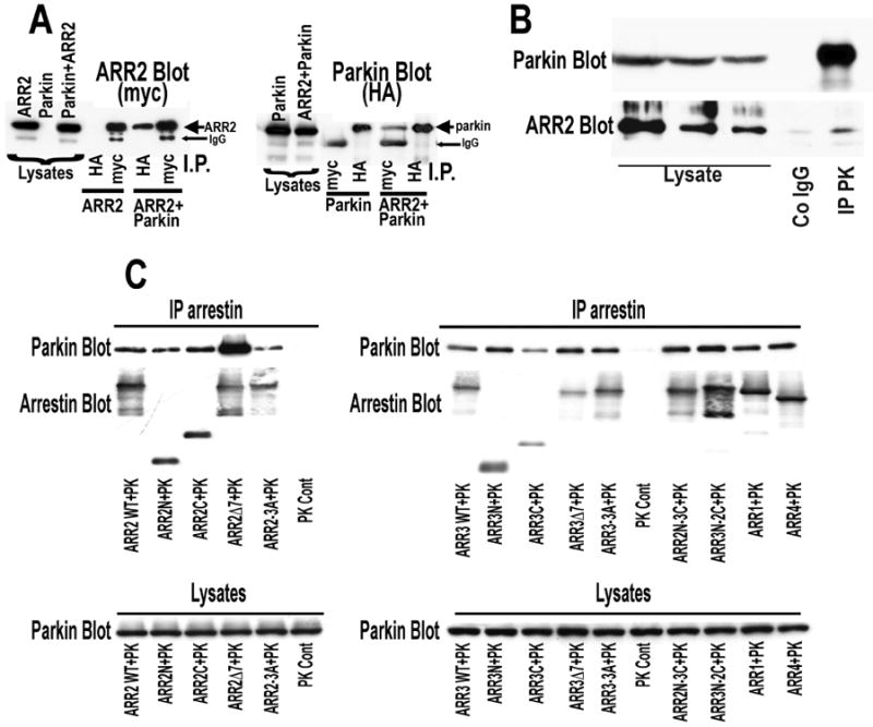
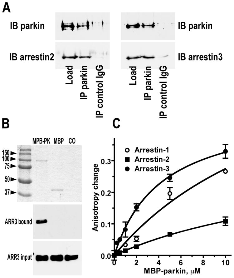
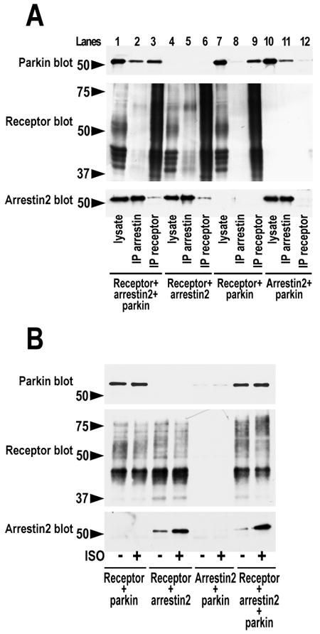
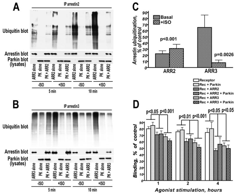
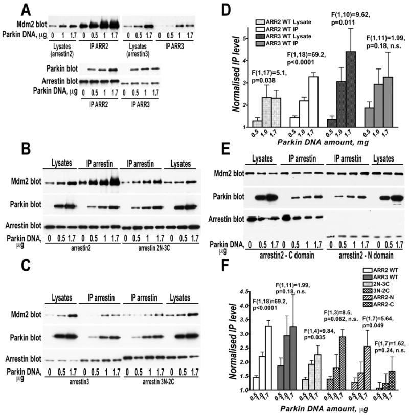
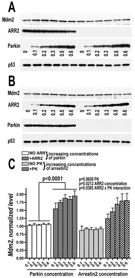
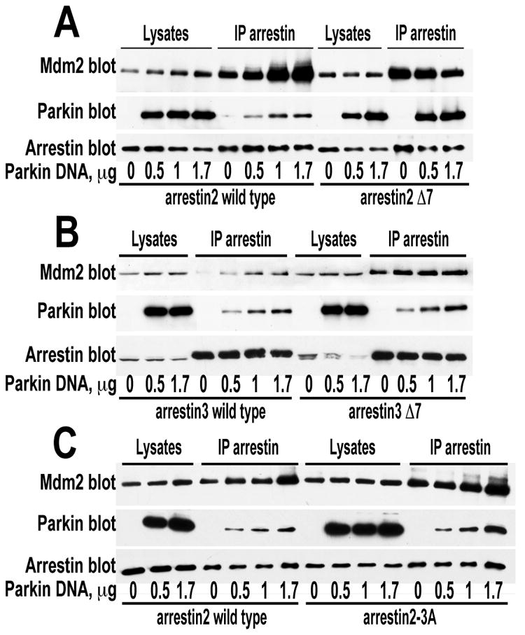
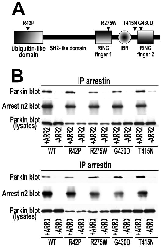
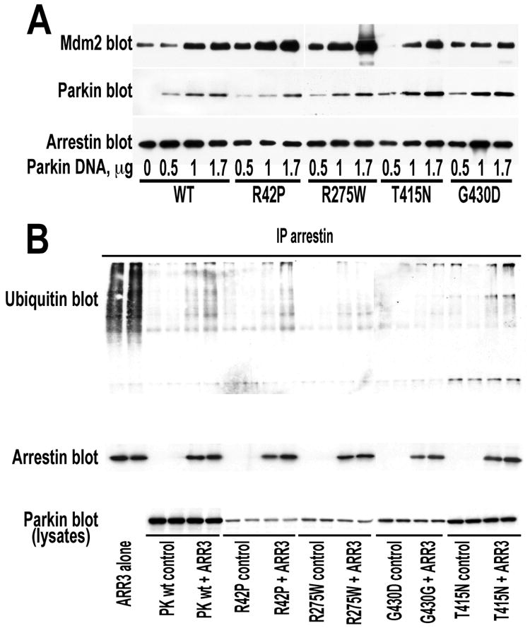
Numerous mutations in E3 ubiquitin ligase parkin were shown to associate with familial Parkinson's disease. Here we show that parkin binds arrestins, versatile regulators of cell signaling. Arrestin-parkin interaction was demonstrated by coimmunoprecipitation of endogenous proteins from brain tissue and shown to be direct using purified proteins. Parkin binding enhances arrestin interactions with another E3 ubiquitin ligase, Mdm2, apparently by shifting arrestin conformational equilibrium to the basal state preferred by Mdm2. Although Mdm2 was reported to ubiquitinate arrestins, parkin-dependent increase in Mdm2 binding dramatically reduces the ubiquitination of both nonvisual arrestins, basal and stimulated by receptor activation, without affecting receptor internalization. Several disease-associated parkin mutations differentially affect the stimulation of Mdm2 binding. All parkin mutants tested effectively suppress arrestin ubiquitination, suggesting that bound parkin shields arrestin lysines targeted by Mdm2. Parkin binding to arrestins along with its effects on arrestin interaction with Mdm2 and ubiquitination is a novel function of this protein with implications for Parkinson's disease pathology.


Figures

Fig. 1
Fig. 1. Parkin interacts with endogenous and expressed arrestins
A. HEK293 cells were transfected with myc-tagged arrestin-2 (ARR2), HA-tagged parkin or both arrestin-2 and parkin. The proteins were immunoprecipitated with anti-HA or anti-myc antibodies as described in methods. Left panel: Western blot for myc detecting arrestin2 in cell lysates and IP products. Right panel: Western blot for HA detecting parkin in cell lysates and IP products. B. Endogenous arrestin-2 (prevalent isoform in the brain) immunoprecipitates with endogenous parkin from rat brain. Parkin (upper blot) and arrestin-2 (lower blot) detected in aliquots of rat cortical lysates and sample immunoprecipitated with anti-parkin antibody (IP PK), but not control IgG (Co IgG). C. Parkin interacts with multiple arrestin isoforms and mutants. Left panel: immunoprecipitation (IP) with anti-FLAG antibodies from HEK293 cells expressing myc-tagged parkin alone (control) or with FLAG-tagged WT and indicated mutant forms of arrestin-2. Right panel: IP with anti-FLAG antibodies from HEK293 cells expressing myc-tagged parkin alone (control) or with FLAG-tagged WT and mutant arrestin-3 (ARR3), and arrestin-2/3 chimeras containing arrestin-2 N-domain and arrestin-3 C-domain (ARR2N-3C), or vise versa (ARR3N-2C), arrestin-1 (ARR1), also known as rod arrestin, and arrestin-4 (ARR4) also known as cone arrestin. Even in the presence of protease inhibitor cocktail some WT and mutant arrestins appear to undergo partial proteolysis during IP generating faster running bands that are not observed in lysates.
Fig. 2
Fig. 2. Parkin binds arrestins directly with micromolar affinity
A. Purified His-parkin (6 μg) was mixed with 6 μg of purified arrestin-2 (left) or -3 (right) and immunoprecipitated overnight (14 h) with anti-parkin (IP parkin) and control (IP control IgG) antibodies (50 μg) covalently attached to 50 μl of the matrix. Aliquots of input and retained proteins were subjected to SDS-PAGE and detected by Western blot with rabbit anti-parkin (IB parkin), anti-arrestin-2 (IB arrestin2), or anti-arrestin-3 (IB arrestin3) antibodies. B. Purified MBP-Parkin (15 μg) (MBP-PK), MBP (15 μg) (MBP), or binding buffer (20 mM Hepes, pH 7.3, 150 mM NaCl)(CO) in 50 μl were loaded onto 50 μl Amylose resin and incubated with purified arrestin-3 (5 μg) at 4°C for 3 h. After three washes bound proteins were eluted with 100 μl of buffer containing 100 mM maltose. The eluted samples were analyzed by SDS-PAGE. Top panel shows Coomassie-stained gel with indicated eluted proteins, middle and bottom panels show Western blots of eluted arrestin-3 (ARR3) and aliquots of the input. C. Indicated purified arrestins (1 μM) labeled with bimane at unique cysteines, as described in methods, were irradiated with polarized 380 nm light. Anisotropy of emitted 470 nm light in the absence and presence of indicated concentrations of purified MBP-parkin is shown (means ± SD from ten measurements in representative experiments). The results of three independent experiments yield apparent KD of MBP-parkin binding to arrestin-1, -2, and -3 of 15.54±10.12 μM, 20.07±5.60 μM, and 3.36±1.86 μM, respectively.
Fig. 3
Fig. 3. Receptor and parkin do not compete for arrestin binding
A. HEK293 cells were transfected with indicated combinations of HA-tagged chimeric β2-adrenergic with the C-terminus of V2 vasopressin receptor, FLAG-tagged arrestin-2, and myc-tagged parkin. Receptor or arrestin was immunoprecipitated with anti-HA (IP receptor) or anti-FLAG (IP arrestin) antibodies, respectively. Parkin (upper blot), receptor (middle blot) and arrestin (lower blot) in aliquots of lysates and immunoprecipitated samples were visualized with appropriate antibodies. B. In the same experimental design, before lysis the cells were incubated for 15 min at 37°C in control medium or with 10 μM of β2-adrenergic agonist isoproterenol (ISO). Receptor was immunoprecipitated with anti-HA antibody, and indicated proteins that co-IP with the receptor were visualized with appropriate antibodies. Whereas the amount of arrestin-2 co-immunoprecipitated with the receptor significantly increases upon receptor stimulation, the amount of parkin does not. The positions of molecular weight markers are indicated on the left. The results of a representative experiments out 2-3 performed are shown.
Fig. 4
Fig. 4. Parkin dramatically reduces basal and receptor-stimulated ubiquitination of arrestins, but does not affect receptor internalization
A, B. HEK293 cells expressing untagged chimeric β2-adrenergic receptor with the C-terminus of vasopressin V2 receptor were co-transfected HA-tagged ubiquitin and FLAG-tagged arrestin-2 (A) or arrestin-3 (B) with or without myc-tagged parkin, as indicated. The cells were treated for indicated time with or without 10 μM of β2-adrenergic agonist isoproterenol (ISO). In immunoprecipitated arrestin samples the amount of arrestin (middle blots) and its ubiquitination (upper blot) was determined by Western blot with appropriate antibody. The amounts of expressed parkin in cell lysates are shown in the lower blots. Note that because multiple forms of arrestin with different numbers of ubiquitin moieties are generated, the main band of non-ubiquitinated protein is by far the most prominent in arrestin blots. C. Quantification of parkin-dependent suppression of arrestin ubiquitination (panels A and B). The ratios of the amount of ubiquitinated arrestin determined with and without receptor activation in the presence or absence of parkin, expressed as % of control (no parkin) are shown. Representative results of one experiment (out of four performed) are shown in panels A and B. Statistical analysis of the data was performed by two-way repeated measure ANOVA with PARKIN as within group factor and ISO stimulation as a between group factor. The effect of PARKIN on the arrestin-2 ubiquitination was significant (F(1,6)=135.2, p<0.0001) as was the effect of ISO (F(1,6)=7.9, p=0.031), whereas the interaction was not significant, indicating that the degree of parkin-induced inhibition of arrestin-2 ubiquitination was independent of receptor stimulation. The effects of PARKIN and ISO on the arrestin-3 ubiquitination were also significant (F(1,6)=57.8, p=0.0003, and 6.67, p=0.042), as was PARKIN × ISO interaction (F(1,6)=18.8, p=0.0049), indicating that PARKIN inhibits activation-induced arrestin-3 ubiquitination significantly stronger. D. Parkin effect on the arrestin-dependent receptor trafficking. HeLa cells were transfected with the chimeric β2V2 receptor, alone or in combination with arrestin-2, arrestin-3, parkin, or both, as indicated. Serum-starved cells were incubated with 10 μM isoproterenol for indicated time. Control cells were incubated without agonist. Cells were then washed 3 × 500 μl in ice-cold TBS, and cell surface receptor was determined by measuring specific binding of cell-impermeable antagonist [3H]CGP-12177 (2nM). The graph shows means±S.E.M. of 6 (for 1 and 2 h) or 2 (for 4 h) independent experiments. Note that parkin co-expression does not affect receptor internalization at any time point. The data for each time point were statistically analyzed by two-way ANOVA with ARRESTIN and PARKIN as main factors. The effects of ARRESTIN were significant (F(1,30)=9.89, p=0.0005; 13.47, p<0.0001; and F(1,6)=9.41, p=0.014, for 1, 2, and 4 h, respectively) for all time points, whereas no significant effect of PARKIN or PARKIN × ARRESTIN interaction was detected. There was no significant difference between arrestin-2 and arrestin-3.
Fig. 5
Fig. 5. Parkin promotes the arrestin interaction with Mdm2
A, B, C, E. Indicated FLAG-tagged arrestins were immunoprecipitated from HEK293 cells co-expressing HA-tagged Mdm2 and varying amounts of WT myc-tagged parkin (the amounts of parkin DNA used for transfection of 60 mm dishes are shown in μg). Upper panels show Western blots for Mdm2; middle panels, parkin; lower panels, arrestins (ARR2, arrestin-2; ARR3, arrestin-3; arrestin-2N-3C, chimera with the N-domain of arrestin-2 and C-domain of arrestin-3; arrestin-3N-2C, reverse chimera; arrestin-2 N- and C-domains included residues 1–180 and 179–418, respectively). Lanes represent equal aliquots of cell lysates or immunoprecipitated samples, as indicated. In panel E, parkin appears to reduce somewhat the expression of the C-domain, but this effect was not studied further. D. Quantification of the effect of parkin concentration on the amount of Mdm2 in cell lysates containing equal amounts of total protein and expressed arrestins, and on the amount of Mdm2 co-immunoprecipitated with equal amount of arrestin-2 or arrestin-3. The data were normalized to the Mdm2 levels in cells or IP samples without parkin. Note that parkin co-expression significantly increases the level of Mdm2 in cells and, to a greater extent, its co-IP with arrestin-2. F. Quantification of parkin effect on the amount of Mdm2 co-immunoprecipitated with equal amount of indicated arrestins. The results of a representative experiments out 2-3 performed are shown. The data were normalized to the Mdm2 level in the IP samples without parkin. The data for each WT and mutant arrestin were analyzed by ANCOVA with Parkin concentration as main factor. The F and p values for each protein are shown on the graphs.
Fig. 6
Fig. 6. Endogenous Mdm2 in the cell is stabilized only by simultaneous presense of arrestin and parkin
A. Cos7 cells were either co-transfected with a fixed amount of FLAG-arrestin2 and increasing amount of myc-parkin or with increasing amount of myc-parkin alone. Representative Western blots show increasing expression of endogenous Mdm2 (upper panel) in cells co-expressing arrestin-2 (middle panel) and parkin (lower panel) but not in cell expressing parkin alone. The data were normalized to the Mdm2 concentration in cells expressing either no parkin (left two sets of panels) or no arrestin-2 (right two sets of panels). Cytosolic p53 served as loading control. B. A reverse experiment to the one shown in A. Cos7 cells were either co-transfected with a fixed amount of myc-parkin and increasing amount of arrestin-2 or with increasing amount of arrestin-2 alone. Representative Western blots show increasing expression of endogenous Mdm2 in cells co-expressing arrestin-2 and parkin but not in cell expressing parkin alone. C. Quantification of the Western blot data demonstrating increased expression of endogenous Mdm2 in cell co-expressing arrestin-2 and parkin. The data are presented as ratios to the values obtained without expressed parkin (experiment shown in A; left side of the graph) or without arrestin-2 (experiment shown in B; right side of the graph). The data were analyzed by ANCOVA with Arrestin or Parkin as a factor and Parkin or Arrestin concentration as a co-variate. The results of 3 independent experiments are shown.
Fig. 7
Fig. 7. Enhanced Mdm2 recruitment by arrestin mutants frozen in the basal state is not increased by parkin
A, B, C. HEK293 cells were co-transfected with HA-Mdm2, indicated FLAG-arrestins, and varying amounts of myc-parkin. Arrestins were immunoprecipitated with anti-FLAg antibodies. Upper blots show Mdm2; middle blots, parkin, lower blots, arrestin. Lanes represent equal aliquots of cell lysates or immunoprecipitated samples, as indicated. Arrestin-2 Δ7 and arrestin-3 Δ7, mutants with seven-residue deletions in the inter-domain hinge, which freezes the protein in the basal conformation and greatly impedes receptor binding; arrestin2-3A, conformationally loose mutant mimicking the active state, that shows enhanced receptor binding. Note that Δ7 mutants of both arrestins bind more Mdm2 than corresponding WT forms, and parkin does not further increase Mdm2 recruitment to these mutants. In contrast, arrestin2-3A mutant shows even more evident enhancement of Mdm2 recruitment by parkin than WT arrestin-2. The results of a representative experiments out 2-3 performed are shown.
Fig. 8
Fig. 8. Parkin mutants implicated in the familial Parkinson's disease bind both non-visual arrestins
A. Domain structure of parkin showing the positions of mutations tested. B. Myc-tagged WT parkin and indicated mutants were expressed in HEK293 cells alone (-ARR2 and –ARR3 controls) or together with FLAG-tagged WT arrestin-2 (+ARR2) or arrestin-3 (+ARR3). Indicated arrestins were immunoprecipitated with anti-FLAG antibody, and IP samples were analyzed by Western blot for parkin (upper blots) and arrestin (middle blots). Lower blots show parkin expression in cell lysates. The results of representative experiments out 2-3 performed with each parkin-arrestin combination are shown.
Fig. 9
Fig. 9. Parkin mutants reduce receptor-stimulated ubiquitination of arrestins and differentially promote Mdm2 recruitment
A. HEK293 cells were co-transfected with HA-Mdm2, FLAG-arrestin-2, and varying amounts of myc-tagged WT parkin or indicated mutants. Arrestin was immunoprecipitated with anti-FLAG antibody, and IP samples were analyzed by Western blot for the presence of Mdm2 (upper blot), parkin (middle blot), and arrestin (lower blot). B. HEK293 cells expressing β2V2 receptor chimera, HA-tagged ubiquitin, myc-tagged parkin mutants alone (control) or with FLAG-tagged WT arrestin-3. Arrestin was immunoprecipitated with anti-FLAG antibody, and IP samples were analyzed by Western blot for the presence of ubiquitin (upper blot) and arrestin (middle blot). The expression of WT parkin and indicated mutants in cell lysates is shown in the lower blot. The results of a representative experiments out 2-3 performed with each arrestin-parkin combination are shown.

References

    1. Kitada T, Asakawa S, Hattori N, Matsumine H, Yamamura Y, Minoshima S, Yokochi M, Mizuno Y, Shimizu N. Mutations in the parkin gene cause autosomal recessive juvenile parkinsonism. Nature. 1998;392:544–545. - PubMed
    1. Lücking CB, Dürr A, Bonifati V, Vaughan J, De Michele G, Gasser T, Harhangi BS, Meco G, Denèfle P, Wood NW, Agid Y, Brice A French Parkinson's Disease Genetics Study Group, and European Consortium on Genetic Susceptibility in Parkinson's Disease. Association between Early-Onset Parkinson's Disease and Mutations in the Parkin Gene. N Engl J Med. 2000;342:1560–1567. - PubMed
    1. Khan NL, Scherfler C, Graham E, Bhatia KP, Quinn N, Lees AJ, Brooks DJ, Wood NW, Piccini P. Dopaminergic dysfunction in unrelated, asymptomatic carriers of a single parkin mutation. Neurology. 2005;64:134–136. - PubMed
    1. Farrer M, Chan P, Chen R, Tan L, Lincoln S, Hernandez D, Forno L, Gwinn-Hardy K, Petrucelli L, Hussey J, Singleton A, Tanner C, Hardy J, Langston W. Lewy bodies and parkinsonism in families with parkin mutations. Ann Neurol. 2001;50:293–300. - PubMed
    1. Pramstaller PP, Scaravilli F, Eskelson C, Pepivani I, Hedrich K, Gonzales-McNeal M, Hilker R, Kramer PL, Klein C. Lewy body Parkinson's disease in a large pedigree with 77 Parkin mutation carriers. Ann Neurol. 2005;58:411–422. - PubMed

Publication types